. 2009 Nov;37(21):7206-18.
doi: 10.1093/nar/gkp790.

DNA cleavage and methylation specificity of the single polypeptide restriction-modification enzyme LlaGI

Affiliations

DNA cleavage and methylation specificity of the single polypeptide restriction-modification enzyme LlaGI

Rachel M Smith et al. Nucleic Acids Res. 2009 Nov.

Abstract

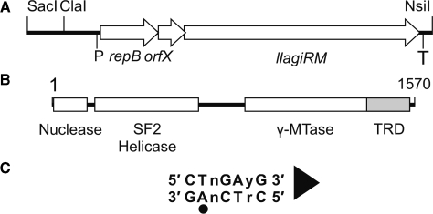
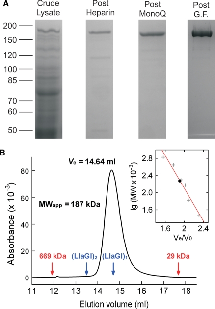
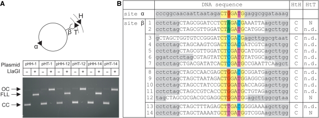
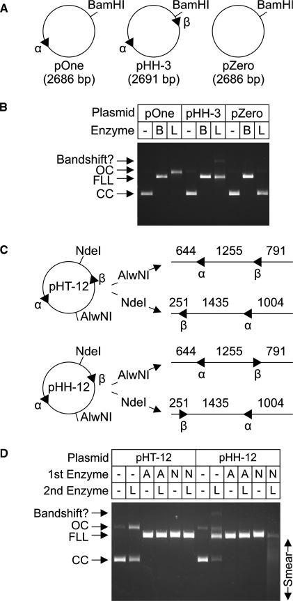
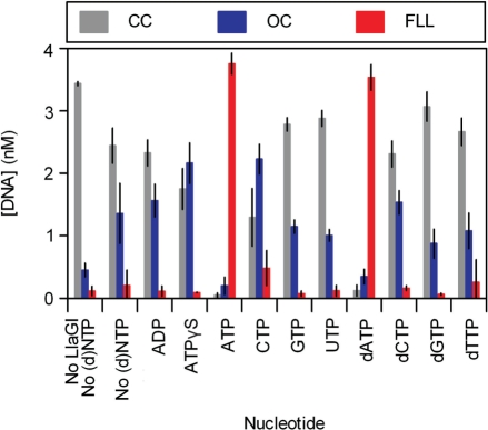
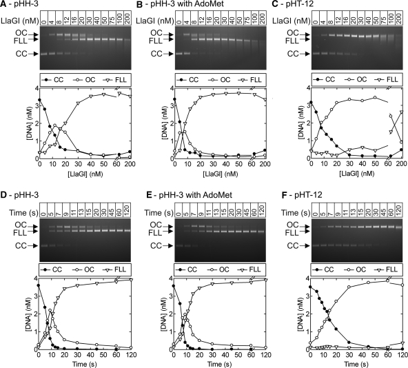
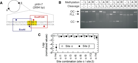
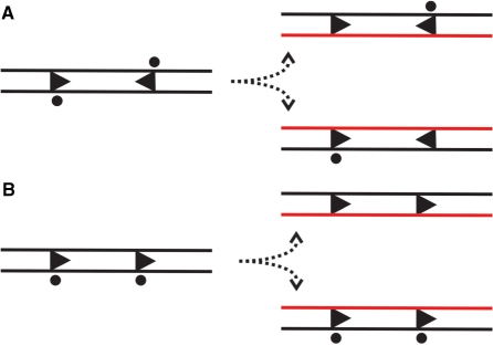
LlaGI is a single polypeptide restriction-modification enzyme encoded on the naturally-occurring plasmid pEW104 isolated from Lactococcus lactis ssp. cremoris W10. Bioinformatics analysis suggests that the enzyme contains domains characteristic of an mrr endonuclease, a superfamily 2 DNA helicase and a gamma-family adenine methyltransferase. LlaGI was expressed and purified from a recombinant clone and its properties characterised. An asymmetric recognition sequence was identified, 5'-CTnGAyG-3' (where n is A, G, C or T and y is C or T). Methylation of the recognition site occurred on only one strand (the non-degenerate dA residue of 5'-CrTCnAG-3' being methylated at the N6 position). Double strand DNA breaks at distant, random sites were only observed when two head-to-head oriented, unmethylated copies of the site were present; single sites or pairs in tail-to-tail or head-to-tail repeat only supported a DNA nicking activity. dsDNA nuclease activity was dependent upon the presence of ATP or dATP. Our results are consistent with a directional long-range communication mechanism that is necessitated by the partial site methylation. In the accompanying manuscript [Smith et al. (2009) The single polypeptide restriction-modification enzyme LlaGI is a self-contained molecular motor that translocates DNA loops], we demonstrate that this communication is via 1-dimensional DNA loop translocation. On the basis of this data and that in the third accompanying manuscript [Smith et al. (2009) An Mrr-family nuclease motif in the single polypeptide restriction-modification enzyme LlaGI], we propose that LlaGI is the prototype of a new sub-classification of Restriction-Modification enzymes, named Type I SP (for Single Polypeptide).


Figures

Figure 1.
Figure 1.
The LlaGI RM enzyme. (A) The SacI-NsiI region of pEW104 identified previously as sufficient for in vivo RM activity (1). The approximate locations of the putative promoter (P) and terminator (T) of the operon are indicated. (B) The llagiRM gene product is a single polypeptide, multi-domain protein (1). Protein domain boundaries were estimated using secondary structure prediction and alignment with LlaBIII (not shown). Target Recognition Domain (TRD). (C) Recognition sequence of LlaGI. The adenine residue that is methylated is indicated by a circle. The arrowhead defines the directionality of the site according to cleavage (Figure 4) and translocation assays (10). The ‘top strand’ sequence (i.e. that starting with CT) is quoted throughout.
Figure 2.
Figure 2.
Purification and solution stoichiometry of LlaGI. (A) Lanes from separate SDS polyacrylamide gels showing stages in the protein purification protocol. G.F., gel filtration. The Post G.F. lane represents 5 µg of purified RM.LlaGI. See ‘Materials and Methods’ section for full details. (B) Elution profile of 263 µg (14.7 µM) LlaGI from a 24 ml Superose 6 column. 100 µl of protein sample was eluted in TMD Buffer plus 15 mM NaCl at 0.4 ml/min. Protein was monitored using absorbance at 280 nm. The elution volumes of the thyroglobulin (669 kDa) and carbonic anhydrase (29 kDa) standards are indicated in red. Indicated in blue are the putative elution volumes of a LlaGI monomer and dimer assuming a theoretical MW of 179 kDa (calculated from the full length amino acid sequence). (Inset) The apparent molecular weight of LlaGI was evaluated from the elution volume using a series of standards (‘Materials and Methods’ section). The red line indicates a least squares linear fit.
Figure 3.
Figure 3.
Cleavage of LlaGI recognition sequences. (A) A library of two-site DNA substrates were generated from pOne (Figure 4) by cloning different LlaGI sequences (Site β – see B). Site α was constant in each case. For three of the sequences both directly-repeated and indirectly-repeated copies of Site β were tested. Example agarose gel shown where CC is Covalently closed Circular substrate DNA, OC is Open Circle nicked intermediate and FLL is the Full Length Linear product cleaved in both strands. DNA and LlaGI were incubated for 10 min. (B) Sequences of sites tested with outcome for indirectly-repeated (HtH) and directly-repeated (HtT) arrangements of sites: C, dsDNA cleavage; N, nicking only; n.d., not determined. pOne sequence is highlighted in grey, cloned sequence in white, constant LlaGI sequence in yellow and degenerate positions in red (dG), green (dA), blue (dC), purple (dT). Sequence shown is the ‘top strand’ as defined in Figure 1C.
Figure 4.
Figure 4.
DNA site requirements for cleavage by LlaGI. (A and B) Plasmid substrates with no sites, one-site or two indirectly-repeated sites were incubated with either saturating BamHI (B) or LlaGI (L) for 1 h. Substrates and products were separated by agarose gel electrophoresis as indicated. (C and D) Plasmid substrates with two directly-repeated sites (pHT-12) or two indirectly-repeated sites (pHH-12) were cleaved with either AlwNI (A) or NdeI (N) to produce the linear DNA indicated. Sequences of the LlaGI sites are in Figure 3. The parental plasmids and linear DNA were then incubated with saturating LlaGI for 1 h. Substrates and products were separated by agarose gel electrophoresis as indicated. See main text for full details. Under these assay conditions, an additional slowly-migrating band was observed which we assign to a LlaGI-DNA bandshift. Gels labelled as in Figure 3.
Figure 5.
Figure 5.
Nucleotide dependence of DNA cleavage by LlaGI. Error bars represent standard errors from at least two repeat experiments. pHH-3 was incubated with excess LlaGI for 10 min in the absence or presence of 4 mM nucleotide, as indicated. Reaction products were separated by agarose gel electrophoresis and the extent of DNA cleavage quantified (‘Materials and Methods’ section).
Figure 6.
Figure 6.
Stoichiometry and rate of DNA cleavage by LlaGI. Points are the mean of at least two repeat experiments. Note that where the FLL product becomes smeared due to end-processing, this can cause an overestimation of the CC band which overlaps with the smear. Similarly, smearing of the OC intermediate in part C causes the CC and FLL bands to be overestimated. This smearing most likely results from processing of the nicked DNA to form ssDNA gaps of different lengths (e.g. by exonucleolytic digestion from the nick). (A) pHH-3 (Figure 3) was incubated with LlaGI at the concentrations indicated for 1 hr. (B) As (A) except in the presence of 100 µM AdoMet. (C) pHT-12 (Figure 3) was incubated with LlaGI at the concentrations indicated for 1 h. (D) Time course of cleavage of pHH-3 with a saturating concentration of LlaGI (200 nM). (E) as (D) except in the presence of 100 µM AdoMet. (F) Time course of cleavage of pHT-12 with a saturating concentration of LlaGI (200 nM). Gels labelled as in Figure 3.
Figure 7.
Figure 7.
Methylation specificity of LlaGI. (A) Plasmid pHH-7 (Figure 3) showing the overlapping EcoAI (blue), LlaGI (bold and yellow) and EcoR124I (red) sequences at Site β. Adenine residues methylated by EcoAI and EcoR124I are indicated by blue and red circles, respectively (2). (B) pHH-7 was treated with LlaGIDA078 (L), M.EcoAI (A) or M.EcoR124I (R), as indicated, in the presence of AdoMet. These DNA substrates were then treated with LlaGI (L), EcoAI (A) or EcoR124I (R), as indicated, in the presence of ATP and AdoMet. The DNA was then separated by agarose gel electrophoresis. See ‘Materials and Methods’ section and main text for full details. Note, cleavage by all three enzymes produces linear DNA that is further processed to generate a DNA smear. (C) pZero, pOne and pHH−1, −2, −3, −6, −8, −9, −13 and −14 (Figure 3) were incubated with LlaGIDA078 and [3H-methyl] AdoMet for 1 h. The extent of 3H-labelling of each site was then assessed by scintillation counting (‘Materials and Methods’ section). For each plasmid, the labelling at each site was corrected for the gel background and the background from the corresponding fragments of pZero. Values were then normalised relative to Site α of pOne. These values are presented as a linear scale where positive values indicate increased labelling relative to Site α and negative values indicate reduced labelling relative to Site α. In pOne the region of Site β is random DNA and therefore acts as a non-specific control. The value of this should tend towards an infinitely small negative value. The observed value of ∼−6 therefore suggests a small background labelling of non-specific sites when a specific site is present on the DNA (i.e. the background labelling is higher than on pZero). In each plasmid the sequence at Site α is the same. Therefore, the values should all be zero, as in plasmid pOne. Given that the values for Sites α and β were very similar on each DNA, small variations between plasmids most likely represent different efficiencies of methylation that arise from variations in DNA preparation quality. Error bars represent the standard error from three repeat experiments.
Figure 8.
Figure 8.
The consequences of recognition site hemi-methylation following replication, adapted from the model of Meisel et al.(32,33). The LlaGI site is indicated as an arrowhead (Figure 1C), the methylated site by a circle and the newly synthesised DNA in red. (A) Where two sites are in head-to-head repeat, one site of the pair is methylated in each of the daughter DNAs and both are protected from dsDNA cleavage (Figure 7). (B) With two sites in head-to-tail repeat, both sites in one daughter DNA are completely unmethylated. However, this arrangement of sites does not result in a dsDNA break (Figures 3 ands 4). DNA nicking that result from the unmethylated sites is likely to be repaired before the replication fork next passes.

References

    1. Madsen A, Josephsen J. The LlaGI restriction and modification system of Lactococcus lactis W10 consists of only one single polypeptide. FEMS Microbiol. Lett. 2001;200:91–96. - PubMed
    1. Roberts RJ, Vincze T, Posfai J, Macelis D. REBASE – enzymes and genes for DNA restriction and modification. Nucleic Acids Res. 2007;35:D269–D270. - PMC - PubMed
    1. Aravind L, Makarova KS, Koonin EV. SURVEY AND SUMMARY: Holliday junction resolvases and related nucleases: identification of new families, phyletic distribution and evolutionary trajectories. Nucleic Acids Res. 2000;28:3417–3432. - PMC - PubMed
    1. Bujnicki JM, Rychlewski L. Identification of a PD-(D/E)XK-like domain with a novel configuration of the endonuclease active site in the methyl-directed restriction enzyme Mrr and its homologs. Gene. 2001;267:183–191. - PubMed
    1. Bourniquel AA, Bickle TA. Complex restriction enzymes: NTP-driven molecular motors. Biochimie. 2002;84:1047–1059. - PubMed

Publication types

MeSH terms

Substances