. 2009 Oct 15;69(20):8208-15.
doi: 10.1158/0008-5472.CAN-09-1132. Epub 2009 Oct 6.

Aldehyde dehydrogenase-expressing colon stem cells contribute to tumorigenesis in the transition from colitis to cancer

Affiliations

Aldehyde dehydrogenase-expressing colon stem cells contribute to tumorigenesis in the transition from colitis to cancer

Joseph E Carpentino et al. Cancer Res. .

Abstract

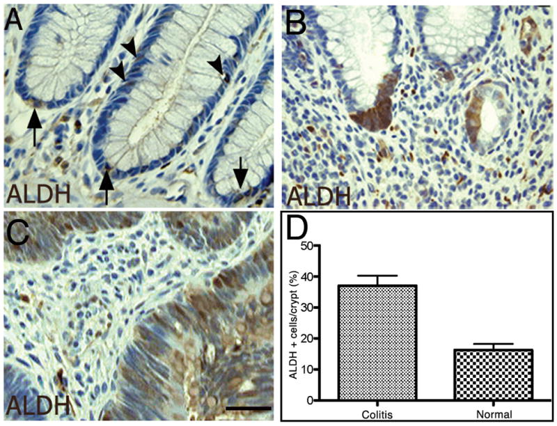
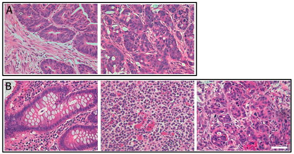
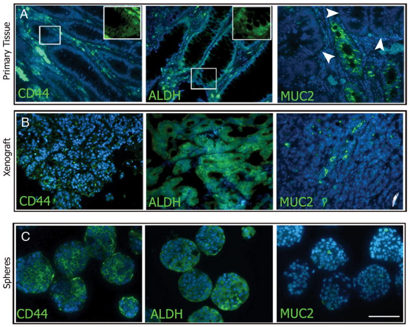
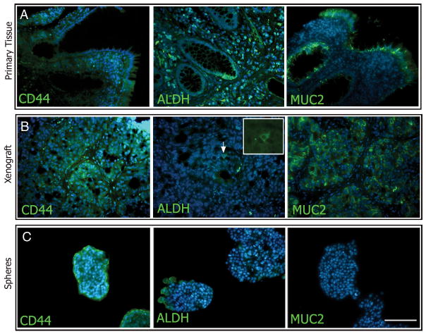
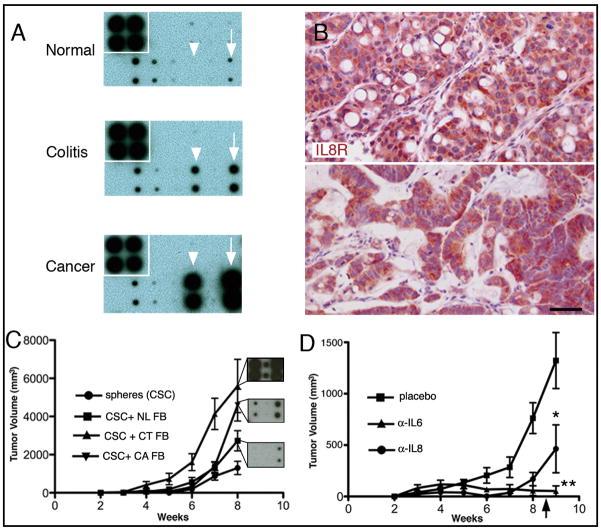
Patients with chronic ulcerative colitis are at increased risk of developing colorectal cancer. Although current hypotheses suggest that sporadic colorectal cancer is due to inability to control cancer stem cells, the cancer stem cell hypothesis has not yet been validated in colitis-associated cancer. Furthermore, the identification of the colitis to cancer transition is challenging. We recently showed that epithelial cells with the increased expression of aldehyde dehydrogenase in sporadic colon cancer correlate closely with tumor-initiating ability. We sought to determine whether ALDH can be used as a marker to isolate tumor-initiating populations from patients with chronic ulcerative colitis. We used fluorescence-activated cell sorting to identify precursor colon cancer stem cells from colitis patients and report both their transition to cancerous stem cells in xenografting studies as well as their ability to generate spheres in vitro. Similar to sporadic colon cancer, these colitis-derived tumors were capable of propagation as sphere cultures. However, unlike the origins of sporadic colon cancer, the primary colitic tissues did not express any histologic evidence of dysplasia. To elucidate a potential mechanism for our findings, we compared the stroma of these different environments and determined that at least one paracrine factor is up-regulated in the inflammatory and malignant stroma compared with resting, normal stroma. These data link colitis and cancer identifying potential tumor-initiating cells from colitic patients, suggesting that sphere and/or xenograft formation will be useful to survey colitic patients at risk of developing cancer.


Figures

Figure 1
Figure 1
Immunohistochemistry of ALDH expression in normal colon, colitis, and colon cancer. Quantification of ALDH+ cells in epithelial tissue. Normal colonic epithelium reveals expression of ALDH at the cells at and near the base of the crypt (A, arrows) with several cells found further up the crypt (arrowheads). The colitic milieu demonstrated expansion of ALDH staining at the base of the crypt (B) compared to baseline normal mucosa. In colitis-associated cancer, the expression of ALDH is greatly expanded (C). ALDH-expressing cells in the epithelial portions of the primary tissue samples reveal an increase in immunoreactive ALDH-expressing cells in the colitic colon (see also Supplementary figure).
Figure 2
Figure 2. Generation of xenografts from colon cancer and colitis
A portion of the colon was removed at operation and tissue was implanted into the flank of NOD-SCID mice. As few as 25 cells of either ESAhighCD44high or ESAhighALDHhigh were capable of reconstituting a tumor and histology strongly resembles the parent tumor (A; N=10). Colitic colon tissue (B, left panel) was implanted into the flank of NOD-SCID mice and observed for xenograft incorporation and growth. As few as 50 ESAhighALDHhigh cells were capable of reconstituting a tumor and H&E staining of tumor revealed anaplastic cells (B, middle panel; N=3). With serial passaging, xenografts from colitic tissues revealed an adenocarcinoma, with the epithelium forming glandlike structures (B, right panel; N=3). Scale Bar = 200 μm.
Figure 3
Figure 3. Marker analysis of primary tissue, xenografts, and spheres from malignant colon tissue
Expression of CD44 and ALDH was abundant in all tissue types examined (A–C left and middle panels, respectively). MUC2 expression in cancerous colon tissue was found in some crypts (A, right panel), while others showed no staining (arrowheads). MUC2 expression was relatively low and sporadic in the xenografts and spheres (B and C right panels, respectively). Scale bar = 200 μm.
Figure 4
Figure 4
Marker analysis of primary tissue, xenografts, and spheres from colitic colons. Expression of CD44 was abundant in all tissue types examined (A–C, left panels). ALDH expression was expanded from the base of the crypts of colitic colons (A, middle panel), were found to be rare in xenografted tissue (B, middle panel), and was found in clusters in colitis-spheres (C, middle panel). MUC2 expression was abundant in the colitic and xenograft tissue (A and B, right panels) but was not observed in colitis-spheres (C, right panel). Scale bar = 200 μm.
Figure 5
Figure 5
Cytokine array analysis of normal, colitic, and cancer stroma. Immunohistochemical expression of CXCR1 (IL8R). Tumorigenicity from cancer derived spheres in the presence/absence of stroma. Cytokine array analysis of conditioned media from normal (A, top), colitic (A, middle), and malignant (A, bottom) human colon. Cytokines IL6 (arrowhead), and IL8 (arrow) are relatively absent in normal conditions (N=4), with increases in colitic (N=3) and malignant conditions (N=3). IL8 receptor expression (CXCR1) in adenocarcinoma generated from colitic tissues reveals widespread expression (B, upper panel, N=2). IL8 receptor expression (CXCR1) in adenocarcinoma originating from human sporadic adenocarcinoma (B, lower panel, N=4). CXCR1 is also widely expressed in xenografts (passage 7) from sporadic colon cancer and resulting tumorigenic growth (B, lower panel). Co-injection of normal, colitic, or cancer-associated fibroblasts reveal a role for stromal elements in tumorigenesis and corresponding cytokine arrays demonstrate a likelihood that IL and/or IL8 play a role (C) p< 0.0001. Insets reveal relative cytokine/chemokine expression for IL6 (left) and IL8 (right). Pellets containing antibodies to IL8 or IL6 resulted in significantly reduced tumor sizes (D) and resulted in persistent inhibition of tumorigenesis until the source of antibodies was exhausted at ~ 8 weeks post-implant (arrow). *p < 0.03; **, p < 0.01; N > or = 4 per group. Scale bar = 100 μm.

References

    1. Kim CF, Dirks PB. Cancer and stem cell biology: how tightly intertwined? Cell Stem Cell. 2008;3:147–50. - PubMed
    1. Dalerba P, Dylla SJ, Park IK, et al. Phenotypic characterization of human colorectal cancer stem cells. Proc Natl Acad Sci U S A. 2007;104:10158–63. - PMC - PubMed
    1. O’Brien CA, Pollett A, Gallinger S, Dick JE. A human colon cancer cell capable of initiating tumour growth in immunodeficient mice. Nature. 2007;445:106–10. - PubMed
    1. Ricci-Vitiani L, Lombardi DG, Pilozzi E, et al. Identification and expansion of human colon-cancer-initiating cells. Nature. 2007;445:111–15. - PubMed
    1. Al-Hajj M, Wicha MS, Benito-Hernandez A, Morrison SJ, Clarke MF. Prospective identification of tumorigenic breast cancer cells. Proc Natl Acad Sci U S A. 2003;100:3983–88. - PMC - PubMed

Publication types

MeSH terms