. 2009 Oct 6;2(91):ra60.
doi: 10.1126/scisignal.2000360.

Wingless promotes proliferative growth in a gradient-independent manner

Affiliations

Wingless promotes proliferative growth in a gradient-independent manner

Luis Alberto Baena-Lopez et al. Sci Signal. .

Abstract

Morphogens form concentration gradients that organize patterns of cells and control growth. It has been suggested that, rather than the intensity of morphogen signaling, it is its gradation that is the relevant modulator of cell proliferation. According to this view, the ability of morphogens to regulate growth during development depends on their graded distributions. Here, we describe an experimental test of this model for Wingless, one of the key organizers of wing development in Drosophila. Maximal Wingless signaling suppresses cellular proliferation. In contrast, we found that moderate and uniform amounts of exogenous Wingless, even in the absence of endogenous Wingless, stimulated proliferative growth. Beyond a few cell diameters from the source, Wingless was relatively constant in abundance and thus provided a homogeneous growth-promoting signal. Although morphogen signaling may act in combination with as yet uncharacterized graded growth-promoting pathways, we suggest that the graded nature of morphogen signaling is not required for proliferation, at least in the developing Drosophila wing, during the main period of growth.


Figures

Fig. 1
Fig. 1
Wingless signalling has a bimodal effect on cell proliferation. (A) nubG4; TubG80ts wing (control) at 29°C. (B) nubG4/UAS-ArmS10; TubG80ts wing at 29°C. (C) Detail from (B). The red arrowhead points to an ectopic bristle. Scale bars represent 0.25 mm. (D) The presence of Hsp23, Sens, and Dlp, detected by immunofluorescence in a nubG4; TubG80ts disc cultured at 29°C (control). (E) Expression of the same proteins in a nubG4/UAS-ArmS10; TubG80ts disc cultured at 29°C. Note the expansion of the areas in which Hsp23 and Sens (markers of the wing margin) are produced and the reduction of the area in which Dlp (a marker of wing blade) is found. The effects were weaker in the central domain of the wing where nubG4 was less abundantly expressed [red arrowhead in (E); see also fig. S1A]. (F to H) Wings from flies of the genotype nubG4; TubG80ts [(F); control] or nubG4/UAS-ArmS10; TubG80 ts [(G) and detail in (H)] raised at 25°C. Red arrowheads point to ectopic bristles (H). (I to K) Wings from flies of the genotype nubG4; TubG80ts [(I); control] and nubG4/UAS-ArmS10; TubG80ts [(J) and detail in (K)] that were raised at 17°C. (L) Surface areas of wings from flies of genotype nubG4; TubG80ts or nubG4/UAS-ArmS10; TubG80ts cultured at the indicated temperatures. The areas measured correspond to those shaded in green in (A). Error bars indicate SD, and statistical significance was assessed with a Student’s t test. **P <0.01, n >10. All of the images shown are representative of more than 10 samples examined.
Fig. 2
Fig. 2
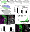
Moderate Wingless signalling stimulates cell proliferation. (A) nubG4 wing from a fly raised at 25°C. (B) nubG4/UAS-WinglessS239A (UASwg[S239A]) wing from a fly raised at 25°C. (C) nubG4 wing from a fly raised at 22°C. (D) nubG4/UAS-Winglessts (UASwg[ts]) wing from a fly raised at 22°C. (E) hhG4 wing from a fly raised at 25°C. Shading marks the A (blue) and P (green) compartments (E). (F) hhG4 UAS-WinglessS239A wing at 25°C. (G) Mean surface area of whole wings (A+P), anterior compartments (A), or posterior compartments (P) in hhG4 (black bars; control) or hhG4 UAS-WinglessS239A (white bars). Statistical significance was as¬sessed with the Student’s t test. *P < 0.05, **P < 0.01; n >10. Error bars indicate the SD. (H) Clonal growth of “equivalent” twin spots [one sister is marked by the absence of GFP (absence of white staining on the right-hand side) whereas the other is marked with 2· GFP, intense white staining on the right-hand side]. These twin spots were generated in an hhG4/UAS-WinglessS239A background. (I) Histogram showing the surface area of 2· GFP clones, located in the anterior (A, control, black bars) or posterior (P, expressing WinglessS239A, green bars) compartments (P < 0.0001, Mann-Whitney test). (J) Nuclear counts in the clone were used to deduce the cell-doubling time (in hours) in the anterior (black bars) and posterior compartments (white bars) of hhG4 (control, left) and hhG4/UAS-WinglessS239A (right) discs. (K) Cell proliferation (as assayed with an antibody specific for PH3, labeled in purple) was relatively increased in the WinglessS239A-expressing posterior compartment. Three representative examples of (K) were recorded with n >10 for all other panels. Scale bars represent 0.5 mm.
Fig. 3
Fig. 3
Wingless signalling promotes cell proliferation in a gradient-independent manner. (A to H) Imaginal discs and wings comprising a posterior compartment composed entirely of wgCX4 homozygous cells (recognized by the absence of GFP). In (E) to (H), all of the cells of the posterior compartment expressed WinglessS239A (hhG4 UAS-WinglessS239A) in addition to being devoid of endogenous Wingless. Wg (A and E), Vg (C and G), and PH3 (D and H) stainings are shown in purple. (I) Histogram showing the ratio between the surface areas of the anterior and posterior compartments in various genetic backgrounds as described below. In the wild type, the A/P ratio was around 1.1. This ratio was slightly changed by the presence of P35 in the posterior compartment (black bar). Lack of Wingless in the posterior compartment [as in (A) to (D)] caused a reduction in surface area, hence an increased A/P ratio (gray bar). This reduction was rescued by adding WinglessS239A [white bar, genotype as in (E) to (H)]. By contrast, P35 had only a mild rescuing effect (purple bar; see also fig. S2, C and D). (J) The surface density of PH3 staining was significantly rescued by the addition of WinglessS239.AStudent’s t test was used to assess significance (**P < 0.01). Error bars indicate the SD. See the Materials and Methods for more details about the genotypes used. Scale bars represent 50 mm for confocal images and 0.5 mm for adult wings. More than 10 samples were analyzed for all panels except (F), which is representative of 8 samples. WgS239A-expressing flies had to be removed manually from the pupal case; they could not close naturally because of leg defects.
Fig. 4
Fig. 4
The abundance of extracellular Wingless determines its effect on growth and the expression of target genes. (A) The amount of extracellular Wingless produced by the UAS-WinglessS239A transgene. The posterior compartment was devoid of endogenous Wingless and expressed UAS-WinglessS239A under the control of hhG4 (right half of the disc). White and blue dashes mark the lines used to generate fluorescence intensity scans. (B) Fluorescent intensity showing the relative amounts of extracellular Wingless in the anterior (A, black line) and posterior (P, blue line) compartments. (C) A disc from a wild-type fly stained at the end of third instar for extracellular Wingless (Extracell. Wg, green), Dally-like (Dlp, red), and Senseless (Sens, blue). White dashes define the line used for the fluorescence intensity measurements shown in (D), which shows the intensity profile for extracellular Wingless (blue line) and Dlp (red line). (E) Diagram correlating the presence of extracellular Wingless with the expression of target genes and the proliferative response. The bracket in (D) and (E) indicates the prospective wing margin. All of the panels are representative of more than 10 samples examined.

References

    1. Ashe HL, Briscoe J. The interpretation of morphogen gradients. Development. 2006;133:385–394. - PubMed
    1. Tabata T, Takei Y. Morphogens, their identification and regulation. Development. 2004;131:703–712. - PubMed
    1. García-Bellido A. Genetic control of wing disc development in Drosophila. Ciba Found. Symp. 1975;0:161–182. - PubMed
    1. Johnston LA, Sanders AL. Wingless promotes cell survival but constrains growth during Drosophila wing development. Nat. Cell Biol. 2003;5:827–833. - PubMed
    1. Herranz H, Milan M. Signalling molecules, growth regulators and cell cycle control in Drosophila. Cell Cycle. 2008;7:3335–3337. - PubMed

Publication types

MeSH terms

LinkOut - more resources