. 2009 Nov;119(11):3450-61.
doi: 10.1172/JCI38432. Epub 2009 Oct 5.

Platelet depletion and aspirin treatment protect mice in a two-event model of transfusion-related acute lung injury

Affiliations

Platelet depletion and aspirin treatment protect mice in a two-event model of transfusion-related acute lung injury

Mark R Looney et al. J Clin Invest. 2009 Nov.

Abstract

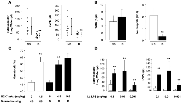
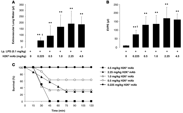
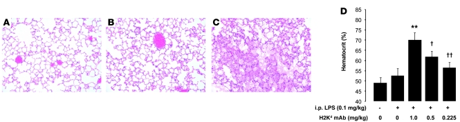
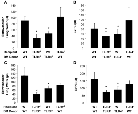
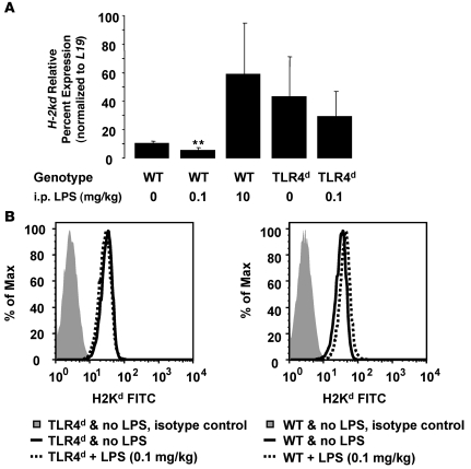
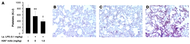
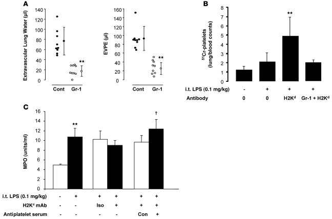
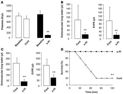
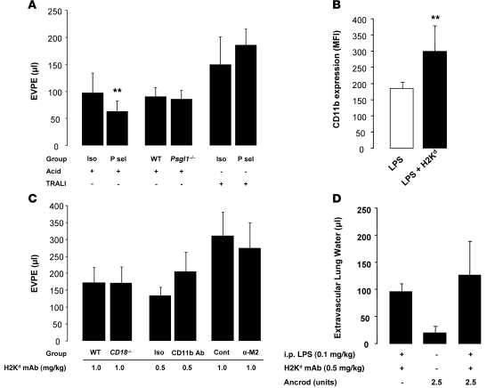
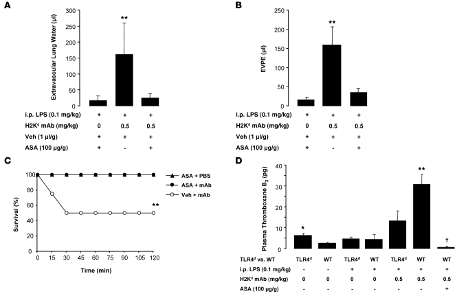
Transfusion-related acute lung injury (TRALI) is the leading cause of transfusion-associated mortality in the US. Previously, we established an immune-mediated TRALI mouse model, wherein mice with cognate antigen were challenged with MHC class I mAb. In this study, when mice housed in a rodent, specific pathogen-free barrier room were challenged with MHC I mAb, there was significant protection from TRALI compared with nonbarrier mice. Priming mice with LPS restored lung injury with mAb challenge. Using TLR4-deficient bone marrow chimeras, the priming phenotype was restricted to animals with WT hematopoietic cells, and depletion of either neutrophils or platelets was protective. Both neutrophils and platelets were sequestered in the lungs of mice with TRALI, and retention of platelets was neutrophil dependent. Interestingly, treatment with aspirin prevented lung injury and mortality, but blocking the P selectin or CD11b/CD18 pathways did not. These data suggest a 2-step mechanism of TRALI: priming of hematopoietic cells, followed by vascular deposition of activated neutrophils and platelets that then mediate the severe lung injury. Furthermore, our data offer an explanation for the increased incidence of TRALI in patients with immune priming conditions, and we suggest what we believe to be a novel therapeutic approach.


Figures

Figure 1
Figure 1. BALB/c mice (H2Kd) housed in a barrier, specific pathogen–free room are protected from TRALI and have lower circulating neutrophils compared with that in nonbarrier animals.
LPS i.t. restores lung injury in barrier animals. (A) Extravascular lung water and EVPEs measured 2 hours after injection of MHC I mAb (4.5 mg/kg, i.v.). Vertical lines indicate mean ± SD. **P < 0.05 barrier (B) vs. nonbarrier mice (NB). (B) Baseline peripheral white blood cell and neutrophil counts in nonbarrier and barrier mice. **P < 0.05 barrier vs. nonbarrier mice. (C) Hematocrit response 2 hours after MHC I mAb. **P < 0.05 nonbarrier mice and barrier mice with MHC I mAb (4.5 mg/kg) vs. mice with no Ab, respectively. (D) Extravascular lung water and EVPEs in barrier mice administered i.t. LPS (white bars) and i.t. LPS plus MHC I mAb (4.5 mg/kg; black bars). **P < 0.05 vs. LPS alone.
Figure 2
Figure 2. Priming with i.p. LPS restores lung injury after MHC I mAb challenge.
(A and B) Extravascular lung water and EVPEs in barrier mice administered i.p. LPS (0.1 mg/kg) with and without increasing doses of MHC I mAb. **P < 0.05 vs. 0 mg/kg H2Kd mAb; P < 0.05 vs. 0.5, 1.0, 2.25, and 4.5 mg/kg H2Kd mAb. (C) Survival curves from mice in A and B. P = 0.02 (Fisher’s exact) for trend of increasing mortality, with increasing dose of MHC I mAb.
Figure 3
Figure 3. LPS i.p. plus MHC I mAb produces severe ALI with hemoconcentration.
(AC) Representative H&E-stained lung sections from (A) a control mouse, (B) a mouse given i.p. LPS (24 hours at 0.1 mg/kg), and (C) a mouse given i.p. LPS (0.1 mg/kg) and MHC I mAb (1.0 mg/kg) at 2 hours after injection. Note intra-alveolar edema fluid in C. Original magnification, ×200. (D) Hematocrit response in barrier mice administered i.p. LPS (0.1 mg/kg) with or without MHC I mAb. **P < 0.05 vs. all other groups, P < 0.05 vs. all other groups, ††P < 0.05 vs. all other groups.
Figure 4
Figure 4. i.p. LPS priming produces peripheral blood neutrophilia, followed by neutropenia and lung neutrophil sequestration, after MHC I mAb challenge.
(A) Peripheral neutrophils in control mice and mice administered i.p. LPS (0.1 mg/kg) with and without MHC I mAb (1.0 mg/kg). **P < 0.05 vs. other groups. (BD) Representative Gr-1 mAb immunostaining in (B) control mouse lungs, (C) mice administered i.p. LPS (at 24 hours after injection; 0.1 mg/kg), and (D) mice administered i.p. LPS (0.1 mg/kg) and MHC I mAb (1.0 mg/kg) at 2 hours after injection. Original magnification, ×400.
Figure 5
Figure 5. WT and TLR4-deficient (TLR4d) bone marrow chimeras reveal that WT TLR4 on hematopoietic cells is critical to the priming phenotype.
(A and B) Extravascular lung water and EVPEs in WT and TLR4d bone marrow chimeras administered i.t. LPS (0.1 mg/kg) and MHC I mAb (4.5 mg/kg). *P < 0.05 vs. WT/WT and TLR4 deficient/WT (recipient/bone marrow donor). (C and D) Extravascular lung water and EVPEs in WT and TLR4-deficient bone marrow chimeras administered i.p. LPS (0.1 mg/kg) and MHC I mAb (1.0 mg/kg). *P < 0.05 vs. WT/WT and TLR4 deficient/WT (recipient/bone marrow donor).
Figure 6
Figure 6. Low-dose LPS priming does not increase lung endothelial or neutrophil MHC I expression.
(A) Relative gene expression results for H-2kd normalized to the control gene, L19, in Tie-2–expressing lung endothelial cells isolated from WT and TLR4d mice administered i.p. LPS (0, 0.1, 10 mg/kg; 24 hours prior to cell isolation). **P < 0.05 vs. all other groups. (B) H2Kd fluorescence intensity in Ly-6G+/CD11b+ peripheral blood cells from WT and TLR4d mice administered no LPS or LPS (0.1 mg/kg; 24 hours prior to cell isolation).
Figure 7
Figure 7. Two-hit TRALI with LPS priming leads to mild thrombocytopenia and sequestration of platelets in the lung microcirculation.
(A) Peripheral blood platelet counts in control mice and mice administered i.p. LPS (0.1 mg/kg) with and without MHC I mAb (1.0 mg/kg). **P < 0.05 vs. other groups, P < 0.05 vs. other groups. (BD) Representative CD41 immunostaining in (B) control mouse lungs, (C) mice administered i.p. LPS (at 24 hours after injection; 0.1 mg/kg), and (D) mice administered i.p. LPS (0.1 mg/kg) and MHC I mAb (1.0 mg/kg) at 2 hours after injection. Original magnification, ×200.
Figure 8
Figure 8. Neutrophil depletion protects mice from 2-event TRALI and from platelet sequestration in the lungs.
Platelet depletion does not affect lung neutrophil sequestration. (A) Extravascular lung water and EVPEs in BALB/c WT mice pretreated with either Gr-1 mAb (250 μg, i.p.) or isotype control (cont) and administered i.t. LPS (0.1 mg/kg), followed by MHC I mAb (4.5 mg/kg). Vertical lines indicate mean ± SD. **P < 0.05 vs. isotype control. (B) 51Cr-platelet counts (whole lung/100 μl blood) in WT control mice and mice given i.t. LPS (0.1 mg/kg) with or without MHC I mAb. The far right bar represents mice pretreated with Gr-1 mAb. **P < 0.05 vs. all other groups. (C) Whole lung MPO (units/ml lung supernatant) measurements. LPS (0.1 mg/kg, i.t.), MHC I mAb (4.5 mg/kg), antiplatelet serum (25 μl, i.p.) were used. Iso, isotype control mAb; Con, control rabbit serum; NS, normal saline. **P < 0.05 vs. normal saline; P < 0.05 vs. control serum.
Figure 9
Figure 9. Platelet depletion protects mice from 2-hit TRALI.
(A) Peripheral blood platelet counts in BALB/c WT mice at baseline and after receiving either control or antiplatelet (α-Pl) serum. **P < 0.05 vs. baseline. (B) Extravascular lung water and EVPEs in mice challenged with i.t. LPS (0.1 mg/kg) and MHC I mAb (4.5 mg/kg) and pretreated with either control or antiplatelet serum. **P < 0.05 vs. control. (C) Extravascular lung water and EVPEs in mice challenged with i.p. LPS (0.1 mg/kg) and MHC I mAb (1.0 mg/kg) and pretreated with either control or antiplatelet serum. **P < 0.05 vs. control. (D) Survival curves from mice in C. **P < 0.05 vs. antiplatelet serum.
Figure 10
Figure 10. Blocking P selectin or MAC-1 fails to protect mice from 2-hit TRALI.
(A) Lung vascular permeability (EVPE) in acid (pH 1.0) and 2-event TRALI (i.p. LPS [0.1 mg/kg] plus MHC I mAb [1.0 mg/kg]) in WT mice pretreated with P selectin (P sel) (30 μg) or isotype control mAbs. **P < 0.05 vs. isotype control (acid). Additionally, WT and Psgl1–/– mice were challenged with i.t. acid. Measurement of extravascular lung water revealed a similar pattern among the groups (data not shown). (B) Flow cytometry staining for CD11b expression (MFI) in Ly-6G+ cells from control mice, i.p. LPS (0.1 mg/kg) and LPS plus MHC I mAb (1.0 mg/kg). **P < 0.05 vs. other groups. (C) Lung vascular permeability (EVPE) in 2-event TRALI (i.p. LPS [0.1 mg/kg] plus MHC I mAb [0.5–1.0 mg/kg]) in WT and Cd18–/– mice and WT mice pretreated with CD11b (1 mg/kg), α-M2 (100 μg), and control Abs. (D) Extravascular lung water in WT mice administered Ancrod (2.5 units i.p., 24 hours and 2 hours prior to H2Kd mAb) to deplete fibrinogen and then challenged with LPS plus MHC I mAb.
Figure 11
Figure 11. Treatment with aspirin prevents TRALI.
(AC) Extravascular lung water (A), lung vascular permeability (EVPE) (B), and survival curves (C) in mice treated with aspirin (ASA; 100 μg/g, i.p.) or vehicle (veh) (DMSO; 1 μl/g, i.p.) 30 minutes prior to LPS (0.1 mg/kg, i.p.) and again 2 hours prior to MHC I mAb (0.5 mg/kg) or control (PBS). Data were obtained 2 hours after injection of MHC I mAb or PBS. **P < 0.05 vs. other groups. (D) Plasma thromboxane B2 levels in TLR4d and WT mice challenged with LPS (0.1 mg/kg, i.p.), MHC I mAb (0.5 mg/kg), and ASA (100 μg/g). *P < 0.05 vs. WT plus no LPS; **P < 0.05 vs. all other groups, P < 0.05 vs. all other groups.

References

    1. Toy P., et al. Transfusion-related acute lung injury: definition and review. Crit. Care Med. 2005;33:721–726. doi: 10.1097/01.CCM.0000159849.94750.51. - DOI - PubMed
    1. Holness L., Knippen M.A., Simmons L., Lachenbruch P.A. Fatalities caused by TRALI. Transfus. Med. Rev. 2004;18:184–188. doi: 10.1016/j.tmrv.2004.03.004. - DOI - PubMed
    1. Kopko P.M., Marshall C.S., MacKenzie M.R., Holland P.V., Popovsky M.A. Transfusion-related acute lung injury: report of a clinical look-back investigation. JAMA. 2002;287:1968–1971. doi: 10.1001/jama.287.15.1968. - DOI - PubMed
    1. Looney M.R., Gropper M.A., Matthay M.A. Transfusion-related acute lung injury: a review. Chest. 2004;126:249–258. doi: 10.1378/chest.126.1.249. - DOI - PubMed
    1. Looney M.R., Su X., Van Ziffle J.A., Lowell C.A., Matthay M.A. Neutrophils and their Fc gamma receptors are essential in a mouse model of transfusion-related acute lung injury. J. Clin. Invest. 2006;116:1615–1623. doi: 10.1172/JCI27238. - DOI - PMC - PubMed

Publication types

MeSH terms