. 2009 Nov 3;101(9):1585-95.
doi: 10.1038/sj.bjc.6605308. Epub 2009 Oct 6.

Curcumin induces apoptosis-independent death in oesophageal cancer cells

Affiliations

Curcumin induces apoptosis-independent death in oesophageal cancer cells

G O'Sullivan-Coyne et al. Br J Cancer. .

Abstract

Background: Oesophageal cancer incidence is increasing and survival rates remain extremely poor. Natural agents with potential for chemoprevention include the phytochemical curcumin (diferuloylmethane). We have examined the effects of curcumin on a panel of oesophageal cancer cell lines.

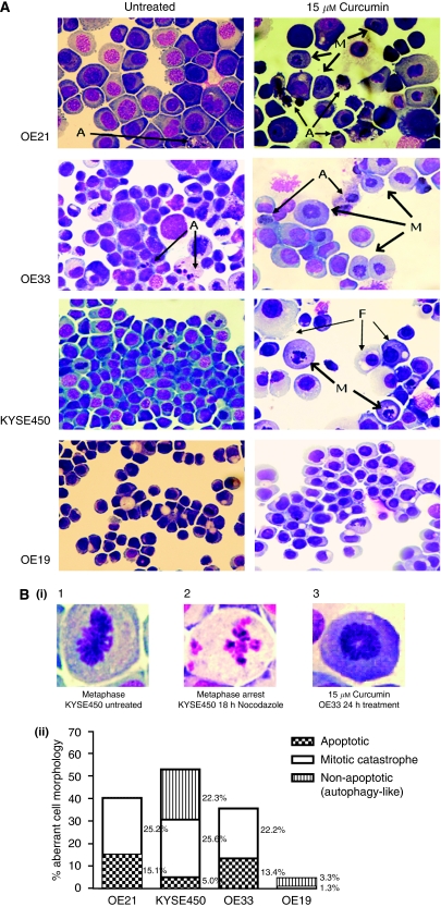
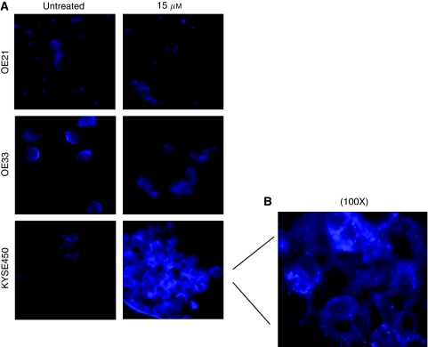
Methods: MTT (3-(4,5-dimethyldiazol-2-yl)-2,5 diphenyl tetrazolium bromide) assays and propidium iodide staining were used to assess viability and DNA content, respectively. Mitotic catastrophe (MC), apoptosis and autophagy were defined by both morphological criteria and markers such as MPM-2, caspase 3 cleavage and monodansylcadaverine (MDC) staining. Cyclin B and poly-ubiquitinated proteins were assessed by western blotting.

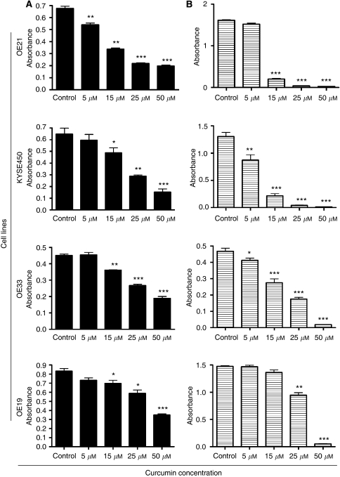
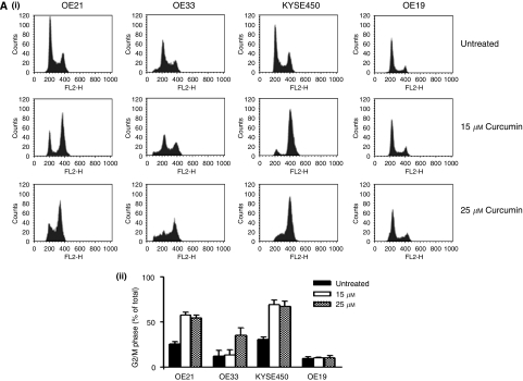
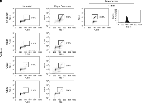
Results: Curcumin treatment reduces viability of all cell lines within 24 h of treatment in a 5-50 muM range. Cytotoxicity is associated with accumulation in G2/M cell-cycle phases and distinct chromatin morphology, consistent with MC. Caspase-3 activation was detected in two out of four cell lines, but was a minor event. The addition of a caspase inhibitor zVAD had a marginal or no effect on cell viability, indicating predominance of a non-apoptotic form of cell death. In two cell lines, features of both MC and autophagy were apparent. Curcumin-responsive cells were found to accumulate poly-ubiquitinated proteins and cyclin B, consistent with a disturbance of the ubiquitin-proteasome system. This effect on a key cell-cycle checkpoint regulator may be responsible for the mitotic disturbances and consequent cytotoxicity of this drug.

Conclusion: Curcumin can induce cell death by a mechanism that is not reliant on apoptosis induction, and thus represents a promising anticancer agent for prevention and treatment of oesophageal cancer.


Figures

Figure 1
Figure 1
Assessment of sensitivity and clonogenicity of squamous cell carcinoma and adenocarcinoma oesophageal cell lines to curcumin. (A) Effects of curcumin on cell viability. After treatment with 5, 15, 25 and 50 μM of curcumin for 24 h, cell viability was determined by MTT assay, with values presented as mean absorbance. *P<0.05, **P<0.005 and ***P<0.0005 compared with untreated cells (paired Student's t-test). Bars=s.e.m. (B) To determine whether or not the cells could overcome the effects of the drug and recover, the curcumin was removed after 24 h of treatment. Cells were allowed to recover for a further 48 h and the MTT assay was repeated. Cell clonogenicity was calculated as the percentage of the control. *P<0.05, **P<0.005 and ***P<0.0005 compared with untreated cells (paired Student's t-test). Bars=s.e.m.
Figure 2
Figure 2
(A) (i) Assessment of DNA content in oesophageal cancer cell lines after 24 h of treatment with curcumin. DNA content analysis was carried out by propidium iodide staining and flow cytometry after 24 h of treatment with increasing concentrations of curcumin. A total of 10 000 cells were counted for each cell line and concentration. Images correspond to typical histogram distributions for each cell line at 0, 15, 25 μM of curcumin. The percentage of cells in the G2/M phase of the cell cycle was estimated using CellQuest software. (ii) The percentage of cells arrested in G2/M phase of the cell cycle. Columns are means of three experiments. Bars=s.d. (B) MPM-2 (anti-phospho-Ser/Thr-Pro) expression in untreated and treated oesophageal cancer cells. MPM-2 is an antibody that recognises a group of proteins that are phosphorylated only in mitosis. Cells were dually stained with propidium iodide to analyse DNA content, and expression was quantified by flow cytometry. As a positive control, KYSE450 cells were treated for 18 h with nocodazole, an anti-fungal agent known to induce metaphase arrest. Cell-cycle analysis and quantification of MPM-2 expression (gated cells) were carried out by flow cytometry after treatment with 25 μM of curcumin for 24 h.
Figure 2
Figure 2
(A) (i) Assessment of DNA content in oesophageal cancer cell lines after 24 h of treatment with curcumin. DNA content analysis was carried out by propidium iodide staining and flow cytometry after 24 h of treatment with increasing concentrations of curcumin. A total of 10 000 cells were counted for each cell line and concentration. Images correspond to typical histogram distributions for each cell line at 0, 15, 25 μM of curcumin. The percentage of cells in the G2/M phase of the cell cycle was estimated using CellQuest software. (ii) The percentage of cells arrested in G2/M phase of the cell cycle. Columns are means of three experiments. Bars=s.d. (B) MPM-2 (anti-phospho-Ser/Thr-Pro) expression in untreated and treated oesophageal cancer cells. MPM-2 is an antibody that recognises a group of proteins that are phosphorylated only in mitosis. Cells were dually stained with propidium iodide to analyse DNA content, and expression was quantified by flow cytometry. As a positive control, KYSE450 cells were treated for 18 h with nocodazole, an anti-fungal agent known to induce metaphase arrest. Cell-cycle analysis and quantification of MPM-2 expression (gated cells) were carried out by flow cytometry after treatment with 25 μM of curcumin for 24 h.
Figure 3
Figure 3
Morphological features of curcumin-induced cell death in oesophageal cancer cell lines. Cell morphology was visualised using RapiDiff staining by light microscopy after treatment with 15 μM of curcumin for 24 h. (A) Typical cytospin images for untreated and curcumin-treated oesophageal cancer cell lines. Curcumin treatment (15 μM) produced a distinct morphology suggestive of a monopolar spindle and duplicated but unseparated chromosomes centrally located in the cell (M); features are consistent with chromatin images described for mitotic catastrophe (MC). In addition to MC, OE21 and OE33 cell lines show a minor population of cells with clear apoptotic morphology (A). Both of these cell lines also have a background of apoptotic cells visible in their untreated cytospins. By contrast, KYSE450 and OE19 cell lines rarely show apoptotic cells. KYSE450-treated cells predominantly show the typical nuclear morphological features of MC after curcumin treatment. Other features coexist at a background level, including nuclear pyknosis, vacuolisation of the cytoplasm or complete loss of the cytoplasmic membrane while the nuclear membrane remains intact; elements consistent morphologically with autophagic cell death (F). OE19-treated cells preserve their overall structural features at this concentration. (B) (i) Typical chromatin organisations in untreated and treated cells. (1) Normal mitosis metaphase image with chromosomes along the equatorial plane of the cell. (2) Nuclear features of cells treated with 18 h of nocodazole, with typical micronucleation. (3) Chromatin alignment in cells treated with curcumin for 24 h. (ii) Distribution of abnormal morphological features in RapiDiff-stained slides in all four oesophageal cancer cell lines after treatment with 15 μM of curcumin over 24 h. Cells were counted on the basis of their morphology (apoptotic, non-apoptotic/MC and autophagy-like) and expressed as the percentage of the total cell population.
Figure 4
Figure 4
Caspase-3 activity and cell viability after treatment with zVAD and curcumin. (A) All four cell lines were treated with 25 μM of curcumin for 24 h. Active caspase-3 activity was estimated by western blotting using equal amounts (40 μg) of whole-cell lysates. β-Actin was used as a loading control. The western blot shown is representative of three independent experiments. (B) Cell viability estimated by MTT assay in all four oesophageal cancer cell lines after treatment with curcumin and pan-caspase inhibitor zVAD-fmk for 24 h. Data was expressed as the percentage of the control. *P<0.05 and **P<0.005 compared with untreated cells (paired Student's t-test). Bars=s.d.
Figure 5
Figure 5
Examination of autophagic cell death in OE21, OE33 and KYSE450 cell lines after treatment with increasing concentrations of curcumin for 24 h. Autophagic vacuoles were identified after incubation of the cells with the selective fluorescent probe monodansylcadaverine (MDC) and visualised immediately by fluorescent microscopy. (A) Fluorescent photographic images of curcumin-sensitive cell lines, OE21, OE33 and KYSE450. Images correspond to untreated and 15 μM of curcumin at 24 h after incubation with MDC. Photographs are representative of two different experiments. Original magnification, × 40. (B) Detailed photograph of the distinct punctate staining in KYSE450 cells after treatment with 15 μM of curcumin and incubation with MDC. Original magnification, × 100.
Figure 6
Figure 6
Poly-ubiquitination and cyclin B1 expression in oesophageal cancer cell lines after treatment with increasing concentrations of curcumin for 24 h. (A) Effects of curcumin treatment on poly-ubiquitination. Equal amounts (30 μg) of whole-cell lysates were separated using SDS–PAGE and protein poly-ubiquitination assessed by western blotting with an anti-ubiquitin antibody. Accumulation of high-molecular-weight ubiquitin complex is evident after curcumin treatment. (B) Cyclin B1 expression in oesophageal squamous and adenocarcinoma cell lines. Equal amounts (30 μg) of whole-cell lysates were separated using SDS–PAGE and protein expression assessed by western blotting with an anti-cyclin B1 antibody. β-Actin was used as a loading control. The western blot shown is representative of three independent experiments.

References

    1. Aggarwal BB, Kumar A, Bharti AC (2003) Anticancer potential of curcumin: preclinical and clinical studies. Anticancer Res 23: 363–398 - PubMed
    1. Aggarwal BB, Shishodia S, Takada Y, Banerjee S, Newman RA, Bueso-Ramos CE, Price JE (2005) Curcumin suppresses the paclitaxel-induced nuclear factor-kappaB pathway in breast cancer cells and inhibits lung metastasis of human breast cancer in nude mice. Clin Cancer Res 11: 7490–7498 - PubMed
    1. Anand P, Kunnumakkara AB, Newman RA, Aggarwal BB (2007) Bioavailability of curcumin: problems and promises. Mol Pharm 4: 807–818 - PubMed
    1. Aoki H, Takada Y, Kondo S, Sawaya R, Aggarwal BB, Kondo Y (2007) Evidence that curcumin suppresses the growth of malignant gliomas in vitro and in vivo through induction of autophagy: role of Akt and extracellular signal-regulated kinase signaling pathways. Mol Pharmacol 72: 29–39 - PubMed
    1. Biederbick A, Kern HF, Elsasser HP (1995) Monodansylcadaverine (MDC) is a specific in vivo marker for autophagic vacuoles. Eur J Cell Biol 66: 3–14 - PubMed

Publication types