. 2009 Oct 7;4(10):e7325.
doi: 10.1371/journal.pone.0007325.

Stem cell therapy with overexpressed VEGF and PDGF genes improves cardiac function in a rat infarct model

Affiliations

Stem cell therapy with overexpressed VEGF and PDGF genes improves cardiac function in a rat infarct model

Hiranmoy Das et al. PLoS One. .

Abstract

Background: Therapeutic potential was evaluated in a rat model of myocardial infarction using nanofiber-expanded human cord blood derived hematopoietic stem cells (CD133+/CD34+) genetically modified with VEGF plus PDGF genes (VIP).

Methods and findings: Myocardial function was monitored every two weeks up to six weeks after therapy. Echocardiography revealed time dependent improvement of left ventricular function evaluated by M-mode, fractional shortening, anterior wall tissue velocity, wall motion score index, strain and strain rate in animals treated with VEGF plus PDGF overexpressed stem cells (VIP) compared to nanofiber expanded cells (Exp), freshly isolated cells (FCB) or media control (Media). Improvement observed was as follows: VIP>Exp> FCB>media. Similar trend was noticed in the exercise capacity of rats on a treadmill. These findings correlated with significantly increased neovascularization in ischemic tissue and markedly reduced infarct area in animals in the VIP group. Stem cells in addition to their usual homing sites such as lung, spleen, bone marrow and liver, also migrated to sites of myocardial ischemia. The improvement of cardiac function correlated with expression of heart tissue connexin 43, a gap junctional protein, and heart tissue angiogenesis related protein molecules like VEGF, pNOS3, NOS2 and GSK3. There was no evidence of upregulation in the molecules of oncogenic potential in genetically modified or other stem cell therapy groups.

Conclusion: Regenerative therapy using nanofiber-expanded hematopoietic stem cells with overexpression of VEGF and PDGF has a favorable impact on the improvement of rat myocardial function accompanied by upregulation of tissue connexin 43 and pro-angiogenic molecules after infarction.


Conflict of interest statement

Competing Interests: Dr. Vincent Pompili has equity interest with Arteriocyte, Inc. Ramasamy Sakthivel is affiliated to the Arteriocyte Inc.

Figures

Figure 1
Figure 1. Evaluation of total ischemic area using Masson's trichrome staining.
Paraffin-blocked tissues from infarct zones were sectioned and stained with Masson's trichrome after six weeks of therapy demonstrating decreased infarct size in VIP < Exp < FCB < Med groups. Five sections were examined from each heart. Four hearts from each group were evaluated. Every section was examined with low power field to visualize gross heart morphology, and to visualize fibrous tissues after ischemia, and area of interest of each heart section is magnified. Representative gross heart pictures are shown here with areas of interest in higher magnification.
Figure 2
Figure 2. Evaluation of cardiac function by echocardiography.
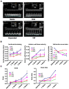
(A) Representative M-mode echocardiographic images after six weeks of therapy demonstrating improved anterior wall motion in the VIP > Exp > FCB > Med groups. Inset shows the short axis two-dimensional image of the LV. (B) Echocardiographic parameters of cardiac function using fractional shortening (FSa), anterior wall tissue velocity (TVa) and wall motion score index (WMSI) plotted as a function of time within each group demonstrating significant improvement in function in the VIP group compared to controls. (C) Strain and strain rates presented as a function of time within each group with significant improvement in the VIP group.
Figure 3
Figure 3. Evaluation of neovascularization.
Cyro-preserved sections were stained with alkaline phosphatase, and were evaluated capillary density area (%) per high power field (A), and total capillary number per high power field (B) in histochemistry images. Representative images of each group (C) were shown in the lower panel. Capillary count and total capillary areas (A & B) were measured by Image Quant software demonstrating highest capillary area density in the VIP group.
Figure 4
Figure 4. Measurement of exercise capacity.
Exercise capacity was evaluated by time on a graded treadmill after six weeks of therapy demonstrated an incremental time in the order of VIP > Exp > FCB > Med groups.
Figure 5
Figure 5. Detection of stem cell homing.
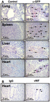
(A) Immunohistochemical detection was performed 36 hours after half a million of GFP overexpressed nanofiber expanded stem cells injected into the left ventricle of the rats, with fixed and paraffin embedded tissue sections from various organs using anti-GFP Ab (right panels) against respective controls (left panels) confirming stem cell homing to lung, spleen, liver and heart. (B) Heart sections were stained for human specific von Willebrand factor (vWF) and immunoglobulin G (IgG) as a control.
Figure 6
Figure 6. Expression of gap junctional protein, connexin 43.
Heart tissue sections were stained for gap junctional protein, connexin 43 from all four groups using immunohistochemistry. Red color indicates connexin 43 stain and blue color indicates DAPI, a nuclear stain. Merged connexin 43 and DAPI of each group is displayed in micrographs.
Figure 7
Figure 7. Expression of signaling molecules.
(A) Western blot of angiogenesis related and connexin 43 proteins from each animal group. (B) Western blot of various molecules of oncogenic potential and relevant signaling pathways from each animal group.

References

    1. Gluckman EG, Roch VV, Chastang C. Use of Cord Blood Cells for Banking and Transplant. Oncologist. 1997;2:340–343. - PubMed
    1. Tse W, Bunting KD, Laughlin MJ. New insights into cord blood stem cell transplantation. Curr Opin Hematol. 2008;15:279–284. - PubMed
    1. Segers VF, Lee RT. Stem-cell therapy for cardiac disease. Nature. 2008;451:937–942. - PubMed
    1. Daley GQ, Scadden DT. Prospects for stem cell-based therapy. Cell. 2008;132:544–548. - PubMed
    1. Singh S, Arora R, Handa K, Khraisat A, Nagajothi N, et al. Stem cells improve left ventricular function in acute myocardial infarction. Clin Cardiol. 2009;32:176–180. - PMC - PubMed

Publication types

MeSH terms