

PAR6, a potential marker for the germ cells selected to form primordial follicles in mouse ovary
- PMID: 19809506
- PMCID: PMC2753645
- DOI: 10.1371/journal.pone.0007372
PAR6, a potential marker for the germ cells selected to form primordial follicles in mouse ovary
Abstract
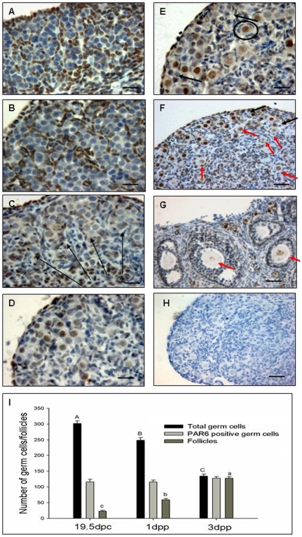
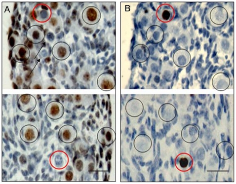
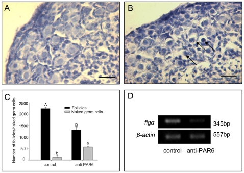
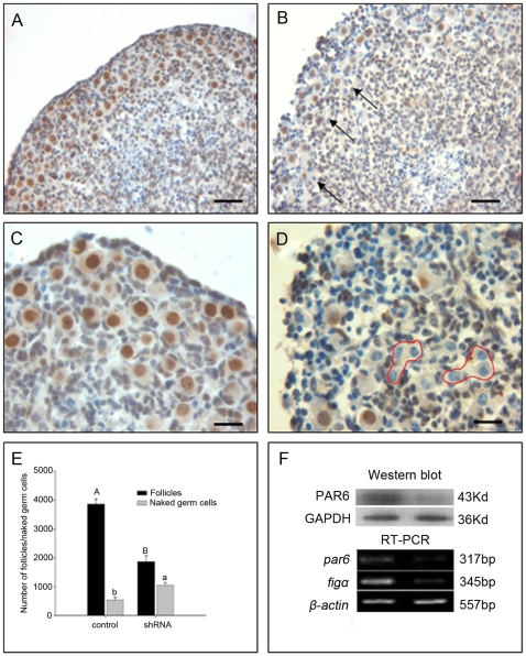
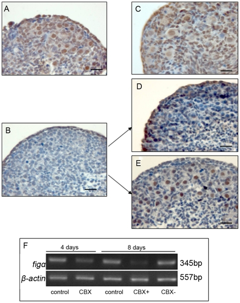
Partitioning-defective proteins (PAR) are detected to express mainly in the cytoplast, and play an important role in cell polarity. However, we showed here that PAR6, one kind of PAR protein, was localized in the nuclei of mouse oocytes that formed primordial follicles during the perinatal period, suggesting a new role of PAR protein. It is the first time we found that, in mouse fetal ovaries, PAR6 appeared in somatic cell cytoplasm and fell weak when somatic cells invaded germ cell cysts at 17.5 days post coitus (dpc). Meanwhile, the expression of PAR6 was observed in cysts, and became strong in the nuclei of some germ cells at 19.5 dpc and all primordial follicular oocytes at 3 day post parturition (dpp), and then obviously declined when the primordial follicles entered the folliculogenic growth phase. During the primordial follicle pool foundation, the number of PAR6 positive germ cells remained steady and was consistent with that of formed follicles at 3 dpp. There were no TUNEL (apoptosis examination) positive germ cells stained with PAR6 at any time studied. The number of follicles significantly declined when 15.5 dpc ovaries were treated with the anti-PAR6 antibody and PAR6 RNA interference. Carbenoxolone (CBX, a known blocker of gap junctions) inhibited the expression of PAR6 in germ cells and the formation of follicles. Our results suggest that PAR6 could be used as a potential marker of germ cells for the primordial follicle formation, and the expression of PAR6 by a gap junction-dependent process may contribute to the formation of primordial follicles and the maintenance of oocytes at the diplotene stage.
Conflict of interest statement
Figures
Similar articles
-
Proliferating cell nuclear antigen (PCNA) regulates primordial follicle assembly by promoting apoptosis of oocytes in fetal and neonatal mouse ovaries.PLoS One. 2011 Jan 6;6(1):e16046. doi: 10.1371/journal.pone.0016046. PLoS One. 2011. PMID: 21253613 Free PMC article.
-
The infant and pubertal human ovary: Balbiani's body-associated VASA expression, immunohistochemical detection of apoptosis-related BCL2 and BAX proteins, and DNA fragmentation.Hum Reprod. 2013 Mar;28(3):698-706. doi: 10.1093/humrep/des453. Epub 2013 Jan 12. Hum Reprod. 2013. PMID: 23315064
-
Arrest at the diplotene stage of meiotic prophase I is delayed by progesterone but is not required for primordial follicle formation in mice.Reprod Biol Endocrinol. 2016 Dec 5;14(1):82. doi: 10.1186/s12958-016-0218-1. Reprod Biol Endocrinol. 2016. PMID: 27919266 Free PMC article.
-
The primordial pool of follicles and nest breakdown in mammalian ovaries.Mol Hum Reprod. 2009 Dec;15(12):795-803. doi: 10.1093/molehr/gap073. Epub 2009 Aug 26. Mol Hum Reprod. 2009. PMID: 19710243 Free PMC article. Review.
-
From primordial germ cells to primordial follicles: a review and visual representation of early ovarian development in mice.J Ovarian Res. 2016 Jun 21;9(1):36. doi: 10.1186/s13048-016-0246-7. J Ovarian Res. 2016. PMID: 27329176 Free PMC article. Review.
Cited by
-
Proliferating cell nuclear antigen (PCNA) regulates primordial follicle assembly by promoting apoptosis of oocytes in fetal and neonatal mouse ovaries.PLoS One. 2011 Jan 6;6(1):e16046. doi: 10.1371/journal.pone.0016046. PLoS One. 2011. PMID: 21253613 Free PMC article.
-
Comparative transcriptome analysis of Indian domestic duck reveals candidate genes associated with egg production.Sci Rep. 2022 Jun 29;12(1):10943. doi: 10.1038/s41598-022-15099-5. Sci Rep. 2022. PMID: 35768515 Free PMC article.
-
HFM1 is essential for the germ cell intercellular bridge transport in primordial follicle formation in mice.Cell Mol Life Sci. 2024 Dec 27;82(1):28. doi: 10.1007/s00018-024-05541-4. Cell Mol Life Sci. 2024. PMID: 39725823 Free PMC article.
-
Construction of conditional acid ceramidase knockout mice and in vivo effects on oocyte development and fertility.Cell Physiol Biochem. 2012;30(3):735-48. doi: 10.1159/000341453. Epub 2012 Aug 1. Cell Physiol Biochem. 2012. PMID: 22854249 Free PMC article.
-
Mechanisms controlling germline cyst breakdown and primordial follicle formation.Cell Mol Life Sci. 2017 Jul;74(14):2547-2566. doi: 10.1007/s00018-017-2480-6. Epub 2017 Feb 14. Cell Mol Life Sci. 2017. PMID: 28197668 Free PMC article. Review.
References
-
- Pepling ME. From primordial germ cell to primordial follicle: Mammalian female germ cell development. Genesis. 2006;44:622–632. - PubMed
-
- Macara IG. Par Proteins: Partners in Polarization. Curr Biol. 2004;14:160–162. - PubMed
-
- Telfer W. Development and physiology of the oocyte-nurse cell syncitium. Adv Insect Physiol. 1975;11:223–319.
-
- Deng W, Lin H. Asymmetric germ cell division and oocyte determination during Drosophila oogenesis. Int Rev Cytol. 2001;203:93–138. - PubMed
Publication types
MeSH terms
Substances
LinkOut - more resources
Full Text Sources
Molecular Biology Databases
Miscellaneous
