. 2009 Sep;41(3):145-54.
doi: 10.4143/crt.2009.41.3.145. Epub 2009 Sep 28.

Apoptosis and expression of AQP5 and TGF-beta in the irradiated rat submandibular gland

Affiliations

Apoptosis and expression of AQP5 and TGF-beta in the irradiated rat submandibular gland

Jin Hwa Choi et al. Cancer Res Treat. 2009 Sep.

Abstract

Purpose: To evaluate the effect of X-ray irradiation on apoptosis and change of expression of aquaporin 5 (AQP5) and transforming growth factor-β(TGF-β) in the rat submandibular gland (SMG).

Materials and methods: SMGs of 120 male Sprague-Dawley rats were irradiated with a single X-ray dose (3, 10, 20, or 30 Gy). At the early and late post-irradiation phase, apoptosis was measured by the terminal deoxynucleotidyl transferase biotin-dUTP nick end labeling (TUNEL) method, and expression of AQP5 and TGF-β was determined by immunohistochemical staining.

Results: At the late post-irradiation phase, increased apoptosis was evident and marked decreases of expression of AQP5 expression by acinar cells and TGF-β expression by ductal cells were evident.

Conclusion: Apoptosis after X-ray irradiation develops relatively late in rat SMG. Irradiation reduces AQP5 and TGF-β expression in different SMG cell types.

Keywords: AQP5; Apoptosis; Radiation; Submandibular gland; TGF-β.


Figures

Fig. 1
Fig. 1
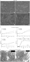
Radiation-induced morphological changes in rat SMG. (A) Nonirradiated gland. The SMG parenchyma is composed mainly of acinar cells and duct cells. (a) Acini, (c) Granular convoluted tubules. (B) Day 3 after irradiation with 10 Gy. Increased numbers of cytoplasmic vacuoles (⇒) in acinar cells were found. (C) Day 60 after irradiation with 10 Gy. Numbers of GCT and SD cells were degenerated and the cells desquamated (→), and some duct cells were dissolved. Sections were stained with H & E. Magnification is ×400.
Fig. 2
Fig. 2
Morphometrical change in GCT cells of irradiated rats at the late post-irradiation phase. *Statistically significant difference from sham-treated control (p<0.05).
Fig. 3
Fig. 3
Apoptosis in submandibular gland of irradiated rats. (A) TUNEL assays in non-irradiated gland (a) and day 3 (b), day 10 (c), and day 30 (d) after irradiation with 10 Gy. (B) Time course of the percentage of apoptotic cells (%) in submandibular gland of irradiated rats. *Statistically significant difference from sham-treated control (p<0.05) (C) The finding in transmission electron microscopy. The apoptotic cells with nuclear chromatin condensation (⇒) and margination (←) were observed at day 30 and 60 after irradiation. Magnification is ×400.
Fig. 4
Fig. 4
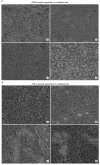
Altered AQP5 and TGF-β expression in irradiated rats. (A) Immunohistochemical staining of AQP5 in non-irradiated gland (a) and day 3 (b), day 10 (c), and day 30 (d) after irradiation with 10 Gy. A loss of AQP5 expression of acinar cells was observed at the late post irradiation phase. (B) Immunohistochemical staining of TGF-β in nonirradiated gland (a) and day 3 (b), day 10 (c), and day 30 (d) after irradiation with 10 Gy. A loss of TGF-β expression of ductal cells was observed at the late post irradiation phase. Magnification is ×400.

References

    1. Taylor JC, Terrell JE, Ronis DL, Fowler KE, Bishop C, Lambert MT, et al. Disability in patients with head and neck cancer. Arch Otolaryngol Head Neck Surg. 2004;130:764–769. - PubMed
    1. Konings AW, Coppes RP, Vissink A. On the mechanism of salivary gland radiosensitivity. Int J Radiat Oncol Biol Phys. 2005;62:1187–1194. - PubMed
    1. Abok K, Brunk U, Jung B, Ericsson J. Morphologic and histochemical studies on the differing radiosensitivity of ductular and acinar cells of the rat submandibular gland. Virchows Arch B Cell Pathol Incl Mol Pathol. 1984;45:443–460. - PubMed
    1. Peter B, Van Waarde MA, Vissink A, s-Gravenmade EJ, Konings AW. The role of secretory granules in the radiosensitivity of rat salivary gland acini--a morphological study. Radiat Res. 1994;140:419–428. - PubMed
    1. Stephens LC, King GK, Peters LJ, Ang KK, Schultheiss TE, Jardine JH. Acute and late radiation injury in rhesus monkey parotid glands. Evidence of interphase cell death. Am J Pathol. 1986;124:469–478. - PMC - PubMed

LinkOut - more resources