. 2009 Dec;3(3-4):255-73.
doi: 10.1007/s12079-009-0072-4. Epub 2009 Oct 7.

SPARC: a matricellular regulator of tumorigenesis

Affiliations

SPARC: a matricellular regulator of tumorigenesis

Shanna A Arnold et al. J Cell Commun Signal. 2009 Dec.

Abstract

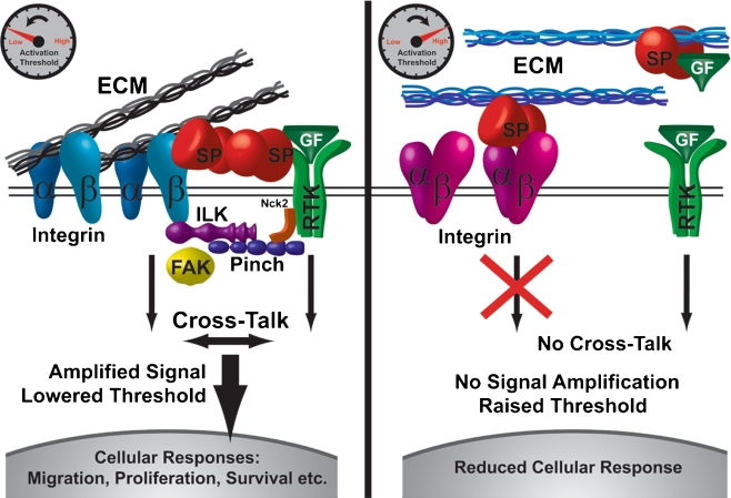
Although many clinical studies have found a correlation of SPARC expression with malignant progression and patient survival, the mechanisms for SPARC function in tumorigenesis and metastasis remain elusive. The activity of SPARC is context- and cell-type-dependent, which is highlighted by the fact that SPARC has shown seemingly contradictory effects on tumor progression in both clinical correlative studies and in animal models. The capacity of SPARC to dictate tumorigenic phenotype has been attributed to its effects on the bioavailability and signaling of integrins and growth factors/chemokines. These molecular pathways contribute to many physiological events affecting malignant progression, including extracellular matrix remodeling, angiogenesis, immune modulation and metastasis. Given that SPARC is credited with such varied activities, this review presents a comprehensive account of the divergent effects of SPARC in human cancers and mouse models, as well as a description of the potential mechanisms by which SPARC mediates these effects. We aim to provide insight into how a matricellular protein such as SPARC might generate paradoxical, yet relevant, tumor outcomes in order to unify an apparently incongruent collection of scientific literature.
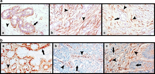

Figures

Fig. 1
Fig. 1
Compartmentalized SPARC expression in human cancer. a Immunohistochemical staining of human biopsies of normal breast and invasive ductal adenocarcinoma, adapted from Barth et al. (2005) Copyright © Springer. Reprinted with permission of Springer-Verlag Berlin Heidelberg New York, A member of BertelsmannSpringer Science + Business Media GmbH (a) SPARC is expressed in myoepithelial cells (arrow) and by a few stromal cells in the ducts of normal breast. However, much of the stroma lacks SPARC expression (arrowhead) (b) Staining for α-SMA in the tumor-associated stroma (arrowheads) reveals activated fibroblasts also positive for (c) SPARC immunoreactivity (arrowheads). The arrow points to malignant epithelial cells lacking SPARC expression. b Immunohistochemical analysis of SPARC expression in colonic mucosae and colorectal carcinomas, adapted from Yang et al. (2007) Copyright © 2005 Wiley-Liss, Inc. Reprinted with permission of Wiley-Liss, Inc., A Wiley Company. (a) Normal colonic epithelial cells (arrow) strongly express SPARC, while there is only minimal SPARC expression in the surrounding stroma (arrowhead). (b, c) SPARC expression by the carcinoma cells (arrows) is dramatically reduced or absent, while tumor stromal cells display strong expression of SPARC (arrowheads) (c) Higher magnification
Fig. 2
Fig. 2
SPARC as an extracellular scaffolding protein and rheostat. We propose that SPARC (SP) acts as an extracellular scaffolding protein; whereby, SPARC controls the interactions and cross-talk between the extracellular matrix (ECM), integrins (α, β) and growth factor receptors (RTK). By controlling integrin clustering and activation, as well as, integrin communication with growth factor receptors, SPARC can function as a rheostat for signaling and cellular response. (Left) SPARC may decrease the activating threshold of certain growth factors (GF) by enhancing complex formation and cross-talk between integrins and growth factor receptors. Integrin-linked kinase (ILK), Pinch, and Nck2 link integrins and growth factor receptors, intracellularly, to form localized signaling cascades, while SPARC acts as an extracellular scaffold to reinforce this complex. Focal adhesion kinase (FAK) is just one example of a signaling molecule located downstream of both integrins and growth factor receptors whose activation is influenced by SPARC. Ultimately, integrin-growth factor receptor cross-talk leads to signal amplification and enhanced cellular responses. (Right) SPARC may also increase the activating threshold of integrins and growth factors by inhibiting the binding of certain integrins to the ECM, opposing integrin-growth factor receptor clustering, and/or sequestering growth factors in the extracellular milieu. All of these effects result in a loss of communication and signal amplification of integrins and growth factor receptors, which reduces cellular responses. ECM composition, integrin profile, cytokine profile, cell-type and SPARC concentration/cell-surface localization are all factors dictating this differential response to SPARC

References

    1. Alonso SR, Tracey L, Ortiz P, Perez-Gomez B, Palacios J, Pollan M, et al. A high-throughput study in melanoma identifies epithelial-mesenchymal transition as a major determinant of metastasis. Cancer Res. 2007;67:3450–3460. - PubMed
    1. Alvarez MJ, Prada F, Salvatierra E, Bravo AI, Lutzky VP, Carbone C, et al. Secreted protein acidic and rich in cysteine produced by human melanoma cells modulates polymorphonuclear leukocyte recruitment and antitumor cytotoxic capacity. Cancer Res. 2005;65:5123–5132. - PubMed
    1. Amatschek S, Koenig U, Auer H, Steinlein P, Pacher M, Gruenfelder A, et al. Tissue-wide expression profiling using cDNA subtraction and microarrays to identify tumor-specific genes. Cancer Res. 2004;64:844–856. - PubMed
    1. Arnold S, Mira E, Muneer S, Korpanty G, Beck AW, Holloway SE, et al. Forced expression of MMP9 rescues the loss of angiogenesis and abrogates metastasis of pancreatic tumors triggered by the absence of host SPARC. Exp Biol Med (Maywood) 2008;233:860–873. - PMC - PubMed
    1. Aycock RL, Bradshaw AC, Sage EH, Starcher B. Development of UV-induced squamous cell carcinomas is suppressed in the absence of SPARC. J Invest Dermatol. 2004;123:592–599. - PubMed