

Chronic surgical site infection due to suture-associated polymicrobial biofilm
- PMID: 19811056
- PMCID: PMC2956523
- DOI: 10.1089/sur.2008.062
Chronic surgical site infection due to suture-associated polymicrobial biofilm
Abstract
Background: Surgical site infection (SSI) is a common surgical complication; culture-negative SSI presents a particular problem in management.
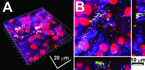
Methods: Examination of explanted foreign bodies (sutures) using confocal laser scanning microscopy (CLSM) and fluorescent in situ hybridization (FISH) after surgical exploration of a chronic culture-negative SSI.
Results: Confocal microscopy (CM) demonstrated bacilli and cocci attached to the surface of the explanted sutures in a mixed biofilm. Fluorescent in situ hybridization confirmed that Staphylococci were components of the mixed biofilm. Removal of the foreign bodies (sutures) resolved the chronic infection.
Conclusion: Chronic SSI can arise from underlying bacterial biofilms, which can invest implanted foreign bodies and associated soft tissue surfaces.
Figures


References
-
- Cheadle WG. Risk factors for surgical site infection. Surg Infect (Larchmt) 2006;7(Suppl 1):S7–S11. - PubMed
-
- Fry DE. The economic costs of surgical site infection. Surg Infect (Larchmt) 2002;3(Suppl 1):S37–S43. - PubMed
-
- Turina M. Cheadle WG. Management of established surgical site infections. Surg Infect (Larchmt) 2006;7(Suppl 3):S33–S41.
-
- Rasnake MS. Dooley DP. Culture-negative surgical site infections. Surg Infect (Larchmt) 2006;7:555–565. - PubMed
-
- Nistico L. Gieseke A. Stoodley P, et al. Fluorescence “in situ” hybridization for the detection of biofilm in the middle ear and upper respiratory tract mucosa. In: Sokolowski B., editor. Auditory and Vestibular Research: Methods and Protocols. Totawa, NJ: Humana Press Inc.; 2008. - PubMed
Publication types
MeSH terms
Grants and funding
LinkOut - more resources
Full Text Sources
