Review
. 2009 Dec;19(6):683-90.
doi: 10.1016/j.sbi.2009.09.001. Epub 2009 Oct 5.

The structural changes of T7 RNA polymerase from transcription initiation to elongation

Affiliations
Review

The structural changes of T7 RNA polymerase from transcription initiation to elongation

Thomas A Steitz. Curr Opin Struct Biol. 2009 Dec.

Abstract

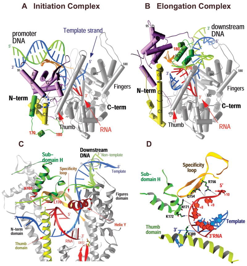
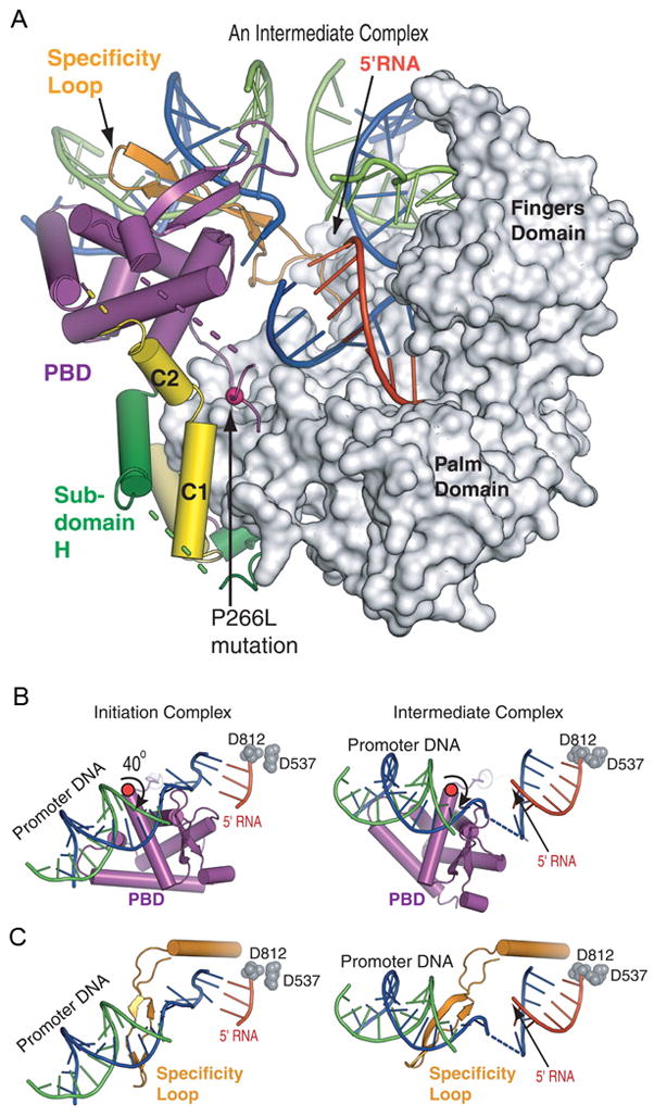
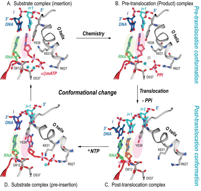
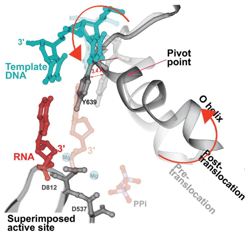
The structures of T7 RNA polymerase (T7 RNAP) captured in the initiation and elongation phases of transcription, as well as an intermediate stage provide insights into how this RNA polymerase protein can initiate RNA synthesis and synthesize 7-10 nucleotides of RNA while remaining bound to the DNA promoter site. Recently, the structures of T7 RNAP bound to its promoter DNA along with either a seven nucleotide or eight nucleotide transcript show an elongated product site resulting from a 40 degrees or 45 degrees rotation of the promoter and domain that binds it. The different functional properties of the initiation and elongation phases of transcription are illuminated from structures of the initiation and elongation complexes. Structural insights into the translocation of the product transcript of RNAP, its separation of the downstream duplex DNA, and its removal of the transcript from the heteroduplex are provided by the structures of several states of nucleotide incorporation. A conformational change in the 'fingers' domain that results from the binding or dissociation of incoming NTP or PPi appears to be associated with the state of translocation of T7 RNAP.


Figures

Figure 1
Figure 1
Comparison of the structures of the T7 RNAP initiation and elongation complexes (A and B) and views of the transcription bubble (C and D). The initiation complex (A) and elongation complex (B) have been orientated equivalently by superimposing their palm domains. Helices are represented by cylinders and beta strands by arrows. The corresponding residues in the NH2-terminal domains of the two complexes that undergo major refolding are colored in yellow, green, and purple, and the COOH-terminal domain (residues 300 to 883) is colored in gray. The template DNA (blue), nontemplate DNA (green), and RNA (red) are represented with ribbon backbones. The proteolysis-susceptible region (residues 170 to 180) is a part of subdomain H (green) in the elongation complex and has moved more than 70 Å from its location in the initiation complex. The specificity loop (brown) recognizes the promoter during initiation and contacts the 5′ end of RNA during elongation, whereas the intercalating hairpin (purple) opens the upstream end of the bubble in the initiation phase and is not involved in elongation. The large conformational change in the NH2-terminal region of T7 RNAP facilitates promoter clearance. This figure was made with the program Ribbons. (C) Interactions of the transcription bubble and heteroduplex in the elongation complex with domain H (green and red) and specificity loop (brown). Proteolytic cuts within the red loop in subdomain H reduce elongation synthesis (21, 22). Thumb alpha-helix (yellow) and alpha-helix Y (orange) are analogously involved in strand separation. (D) Side chains from subdomain H (green), the specificity loop (brown), and the thumb that interact with the single-stranded 5′ end of the RNA transcript and facilitate its separation from the template. The DNA substrate in the initiation complex consisted of duplex promoter DNA from −1 to −17 and a 5′ nucleotide overhang of the template strand (nucleotides 1 to 5) at the 5′ end. The DNA used in the elongation complex contained 10 b.p. of downstream duplex DNA, a 10 nucleotide non-complementary DNA “bubble” and a 10 b.p upstream duplex DNA that is disordered in the structure.
Figure 2
Figure 2
(A) The T7 RNAP in the 7-nt RNA intermediate complex is bound to both promoter and downstream DNA. The PBD has rotated by 40° away from the C-terminal domain, avoiding a steric clash with the transcript and allowing for 7 bp of heteroduplex to form in the active site. (B, C) Promoter and PBD movements during the transition. (B) A view looking down onto the promoter bound to the PBD. A 40° rotation of the PBD away from the active site occurs around an axis that passes through a flexible loop of residues 198 to 204. The catalytic aspartic acid residues (D812 and D537) represent the active site. (C) The same view as in (B) without the PBD but showing the specificity loop, which also rotates.
Figure 3
Figure 3
Structural changes at the active site of T7 RNAP during a single nucleotide addition cycle. This figure shows the O helix with its phosphate binding K631 and R627, a β turn-β motif bearing the metal binding catalytic D812 and D537, template nucleotides in blue, the RNA primer terminus in green, as well as the P and N sites in green and pink ovals. (A) The NTP (red) is bound to the N site in position to be inserted with its metal bound triphosphate moiety crosslinking the O helix to the active site aspartic acid residues. Template nucleotide i+1 (light blue) forms a base pair with the correct incoming nucleotide. (B) The product of the phosphoryl transfer reaction shows a Mg ion (blue) bound to PPi (red), which crosslinks D537 to R627, thereby maintaining RNAP in an identical conformation as in the substrate complex. The 3′ end of RNA remains in the N site in a pre-translocation state. (C) Release of Mg-PPi results in the loss of the link between the O helix and D537, which promotes the rotation of the O helix and translocation of the 3′ end of the RNA to the P site. The RNAP conformational change also places Y639 into the N site and positions the i+2 template nucleotide into the flipped-out, pre-insertion position. (D) A modeled NTP preinsertion complex with NTP bound to the post-translocated RNAP. Although the base binding site is blocked by the side chain of Y639, the triphosphate binding site on the O helix is accessible.
Figure 4
Figure 4
A superposition of the pre- and post-translocation structures at the active site showing the pivoted rotation undergone by the O helix that is associated with translocation (Yin & Steitz, 2004). In the pre-translocation complex (lighter colors), the O helix (light gray) is positioned in the closed conformation by PPi (light red), which is bound to the catalytic carboxyls through Mg. In this conformation, Y639 allows formation of the new base pair (light red and blue). After PPi release, the O helix rotates around Val634, which results in the positive end of the helix moving away from the active site while the other end of the helix moves Y639 by 3.4 Å into the position of the newly formed primer terminus resulting in translocation.

Similar articles

Cited by

References

    1. Cheetham GMT, Steitz TA. Structure of a transcribing T7 RNA polymerase initiation complex. Science. 1999;286:2305. - PubMed
    1. Yin YW, Steitz TA. Structural basis for the transition from initiation to elongation transcription in T7 RNA polymerase. Science. 2002;298:1387. - PubMed
    1. Tahirov TH, Temiakov D, Anikin M, Patlan V, McAllister WT, Vassylyev DG, Yokoyoma S. Structure of a T7 RNA polymerase elongation complex at 2.9 Å resolution. Nature. 2002;420:43. - PubMed
    1. Durniak K, Bailey S, Steitz TA. The structure of a transcribing T7 RNA polymerase in transition from initiation to elongation. Science. 2008;322:553–557. The crystal structures of T7 RNAP bound to substrate DNA and either a 7 nt or 8nt transcript are described. They show a 40° and 45° rotation of the promoter binding domains resulting in an enlargement of the heteroduplex product binding cleft. They represent intermediate states in the transition from the initiation to elongation states. - PMC - PubMed
    1. Yin YW, Steitz TA. The structural mechanism of translocation and helicase activity in T7 RNA polymerase. Cell. 2004;116:393. - PubMed

Publication types

MeSH terms

Substances

LinkOut - more resources