. 2009 Dec;83(24):12822-32.
doi: 10.1128/JVI.01744-09. Epub 2009 Oct 7.

Characterization of a newly identified 35-amino-acid component of the vaccinia virus entry/fusion complex conserved in all chordopoxviruses

Affiliations

Characterization of a newly identified 35-amino-acid component of the vaccinia virus entry/fusion complex conserved in all chordopoxviruses

P S Satheshkumar et al. J Virol. 2009 Dec.

Abstract

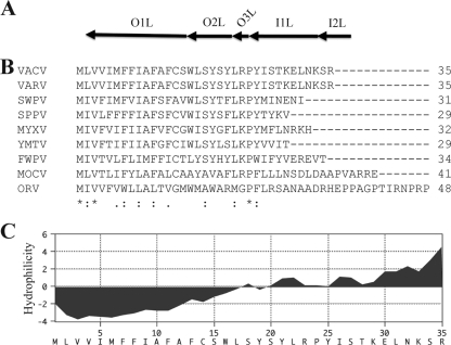
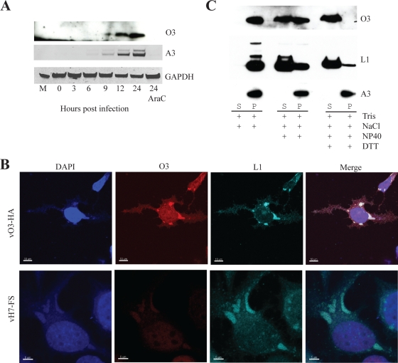
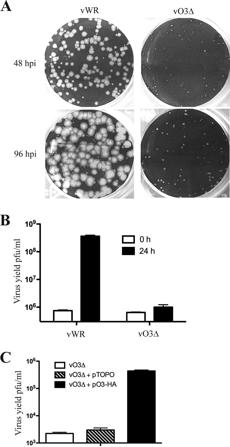
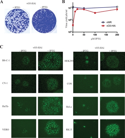
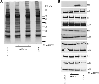
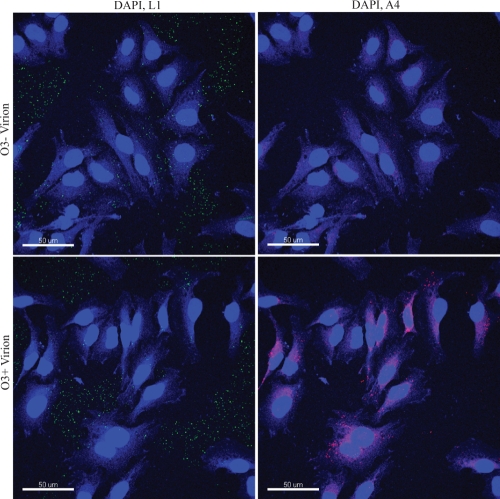
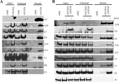
The original annotation of the vaccinia virus (VACV) genome was limited to open reading frames (ORFs) of at least 65 amino acids. Here, we characterized a 35-amino-acid ORF (O3L) located between ORFs O2L and I1L. ORFs similar in length to O3L were found at the same genetic locus in all vertebrate poxviruses. Although amino acid identities were low, the presence of a characteristic N-terminal hydrophobic domain strongly suggested that the other poxvirus genes were orthologs. Further studies demonstrated that the O3 protein was expressed at late times after infection and incorporated into the membrane of the mature virion. An O3L deletion mutant was barely viable, producing tiny plaques and a 3-log reduction in infectious progeny. A mutant VACV with a regulated O3L gene had a similar phenotype in the absence of inducer. There was no apparent defect in virus morphogenesis, though O3-deficient virus had low infectivity. The impairment was shown to be at the stage of virus entry, as cores were not detected in the cytoplasm after virus adsorption. Furthermore, O3-deficient virus did not induce fusion of infected cells when triggered by low pH. These characteristics are hallmarks of a group of proteins that form the entry/fusion complex (EFC). Affinity purification experiments demonstrated an association of O3 with EFC proteins. In addition, the assembly or stability of the EFC was impaired when expression of O3 was repressed. Thus, O3 is the newest recognized component of the EFC and the smallest VACV protein shown to have a function.


Figures

FIG. 1.
FIG. 1.
Computational analysis of the O3L ORF. (A) Diagram of the VACV WR genome indicating ORFs from O1L to I2L. (B) Clustal alignment of VACV O3L ORF with orthologs from representative members of each genus of the Chordopoxvirinae. VACV, vaccinia virus strain WR; VARV, variola virus strain Bangladesh 1975 v75-55-Banu; SWPV, swinepox virus strain Nebraska 17077-99; SPPV, sheeppox virus strain NISKHI; MYXV, myxoma virus strain Lausanne; YMTV, Yaba monkey tumor virus strain Amano; FWPV, fowlpox virus strain Iowa; MOCV, molluscum contagiosum virus strain subtype 1; ORV, Orf virus strain OV-SA00. Amino acid residues conserved in all viruses are denoted by asterisks, those conserved in all but one virus are denoted by colons, and similar amino acids are denoted by periods. (B) Kyte/Doolittle hydrophilicity plot of O3 amino acid sequence analyzed using MacVector 10.6.0 software.
FIG. 2.
FIG. 2.
Temporal expression and localization of O3. (A) Kinetics of expression of O3. BS-C-1 cells were infected with vO3-HA, harvested at the indicated times, and analyzed by SDS-PAGE and Western blotting with anti-HA antibody. Lysates of cells infected in the presence of AraC and harvested at 24 h were analyzed on the same gel. The blot was stripped and probed with antibody to the A3 protein and glyceraldehyde-3-phosphate dehydrogenase (GAPDH) as a loading control. Proteins were detected by chemiluminescence. (B) Confocal microscopy. HeLa cells grown on coverslips were infected with either vO3-HA or control vH7-FS (36). After 18 h, the cells were stained with antibodies against L1 and the HA epitope tag on O3 and corresponding fluorescent dye-conjugated secondary antibodies. DAPI, 4′,6-diamidino-2-phenylindole. (C) Association of O3 with purified virions. MVs were purified from cells infected with vO3-HA, incubated at 37°C with buffers containing the indicated components, and centrifuged to obtain soluble (S) pellet (P) fractions. The pellets were resuspended in the same volume as the soluble fraction, and equivalent amounts were analyzed by SDS-PAGE and Western blotting as in panel A. DTT, dithiothreitol.
FIG. 3.
FIG. 3.
Characterization of the O3L deletion mutant. (A) Plaque phenotypes of VACV WR and vO3Δ. BS-C-1 monolayers were infected with either VACV WR (vWR) or vO3Δ and incubated for 48 or 96 h (postinfection [hpi]) with a methylcellulose overlay and stained with crystal violet to visualize the plaques. (B) Yields of VACV WR and vO3Δ. RK13 cells were infected with VACV WR and vO3Δ at 3 PFU per cell. After 1 h of adsorption, cells were washed to remove the inoculum and harvested immediately or 24 h postinfection. Virus titers were determined by plaque assay in BS-C-1 cells. (C) Transcomplementation of vO3Δ. BS-C-1 cells were infected with vO3Δ and transfected with either the empty vector or plasmid expressing O3-HA under the control of the natural O3L promoter. Cells were harvested after 24 h, and the virus titers were determined by plaque assay.
FIG. 4.
FIG. 4.
Characterization of the O3 inducible mutant virus. (A) Plaque phenotypes of vO3-HAi virus. BS-C-1 monolayers were infected with vO3-HAi and incubated in the absence or presence of 50 μM IPTG with a methylcellulose overlay for 48 h. Plaques were visualized by staining with crystal violet. (B) IPTG dependence of vO3-HAi replication. BS-C-1 cells were infected with 3 PFU per cell of either VACV WR (vWR) or vO3-HAi and incubated with the indicated concentrations of IPTG. After 24 h, the virus yields were determined by plaque assay. (C) Plaque size of vO3-HAi in different cell lines. Confluent cells were infected with vO3-HAi in the absence or presence of inducer for 24 h. Plates were viewed with a fluorescence microscope to show GFP expression, and plaques of a typical size are shown. Cell lines are indicated on the left.
FIG. 5.
FIG. 5.
Proteolytic processing and morphogenesis of VACV in the absence of O3 expression. (A) Proteolytic processing of membrane and core proteins. BS-C-1 cells were infected with vO3-HAi in the presence or absence of 50 μM IPTG, the parental virus vT7LacOI, or vO3Δ. After 24 h, lysates were prepared and analyzed by SDS-PAGE and Western blotting with antibodies to the HA tag on O3, the membrane protein A17, the core protein A3, and glyceraldehyde-3-phosphate dehydrogenase (GAPDH) as a loading control. Bands were visualized by chemiluminescence. (B) Electron microscopy of BS-C-1 cells infected with vO3-HAi in the absence of IPTG. After 20 h, ultrathin cell sections were prepared for viewing with a transmission electron microscope. IV, immature virions; WV, wrapped virions.
FIG. 6.
FIG. 6.
Protein composition of O3 mutant virions. (A) SDS-PAGE. Virions were purified from cells infected with vT7LacOI, vO3Δ, and vO3-HAi in the presence and absence of IPTG; equal numbers of particles, determined by OD, were analyzed by SDS-PAGE. Bands were visualized by silver staining. The positions and masses of marker proteins are shown on the right. (B) Western blotting. Proteins from purified virions were resolved by SDS-PAGE as in panel A and analyzed by Western blotting. The protein targets of the antibodies are indicated on the right.
FIG. 7.
FIG. 7.
Requirement of O3 for syncytium formation. (A) Fusion from within. BS-C-1 cells were infected with vO3-HAi in the absence and presence of IPTG for 18 h at 37°C. The cells were exposed to pH 5.5 or pH 7 buffer (not shown) for 2 min and incubated for an additional 3 h at 37°C in the presence of regular medium. The cells were fixed, stained with DAPI (4′,6-diamidino-2-phenylindole), and examined by phase-contrast and fluorescence microscopy to visualize DAPI and GFP. (B) Fusion from without. BS-C-1 cells were infected with vO3-HAi in the absence and presence of IPTG, and O3 and O3+ virions were purified, respectively. BS-C-1 cells on coverslips were incubated with 200 PFU per cell of O3 or O3+ virions for 1 h at 4°C, exposed to pH 5.5 buffer for 2 min, and incubated for 3 h in regular medium containing 300 μg/ml of cycloheximide at 37°C. The cells were fixed, stained with DAPI and phalloidin (Alexa fluor 594; fluorescent conjugated), and examined by fluorescence microscopy.
FIG. 8.
FIG. 8.
Requirement of O3 for cell entry. O3 and O3+ virions, prepared as in Fig. 7B, were adsorbed to HeLa cells for 1 h at 4°C. The cells were washed and incubated for additional 2 h at 37°C in the presence of medium containing 300 μg/ml of cycloheximide. The cells were fixed, permeabilized, and stained with mouse anti-L1 (green) and rabbit anti-A4 (red) antibodies followed by fluorescent-tagged corresponding secondary antibodies. The images were obtained by confocal microscopy and processed with Imaris software.
FIG. 9.
FIG. 9.
Interaction of O3 with EFC proteins. (A) IP of proteins associated with O3-HA. HeLa cells were infected with 5 PFU per cell of VACV WR (vWR) or vO3-HA. After 24 h, the cells were lysed and incubated with agarose beads coupled to anti-HA antibody. The input, unbound, and eluted bound proteins were resolved by SDS-PAGE, transferred to nitrocellulose membrane, and probed with antibodies to the proteins listed on the right. (B) Association of EFC proteins in the absence of O3. HeLa cells were infected with VACV WR and vO3-HAi-A28TAP with (+) or without (−) IPTG. After 24 h, the cells were lysed and incubated with agarose beads coupled to streptavidin. The input, unbound, and bound proteins associated with A28TAP were analyzed by Western blotting using antibodies to proteins indicated on the right.

References

    1. Alexander, W. A., B. Moss, and T. R. Fuerst. 1992. Regulated expression of foreign genes in vaccinia virus under the control of bacteriophage T7 RNA polymerase and the Escherichia coli lac repressor. J. Virol. 66:2934-2942. - PMC - PubMed
    1. Ansarah-Sobrinho, C., and B. Moss. 2004. Role of the I7 protein in proteolytic processing of vaccinia virus membrane and core components. J. Virol. 78:6335-6343. - PMC - PubMed
    1. Bisht, H., A. S. Weisberg, and B. Moss. 2008. Vaccinia virus L1 protein is required for cell entry and membrane fusion. J. Virol. 82:8687-8694. - PMC - PubMed
    1. Brown, E., T. G. Senkevich, and B. Moss. 2006. Vaccinia virus F9 virion membrane protein is required for entry but not virus assembly, in contrast to the related L1 protein. J. Virol. 80:9455-9464. - PMC - PubMed
    1. Byrd, C. M., and D. E. Hruby. 2005. A conditional-lethal vaccinia virus mutant demonstrates that the I7L gene product is required for virion morphogenesis. Virol. J. 2:4. - PMC - PubMed

Publication types

LinkOut - more resources