. 2009 Nov 1;183(9):6021-9.
doi: 10.4049/jimmunol.0803872. Epub 2009 Oct 7.

Deficiency of type I IFN receptor in lupus-prone New Zealand mixed 2328 mice decreases dendritic cell numbers and activation and protects from disease

Affiliations

Deficiency of type I IFN receptor in lupus-prone New Zealand mixed 2328 mice decreases dendritic cell numbers and activation and protects from disease

Hemant Agrawal et al. J Immunol. .

Abstract

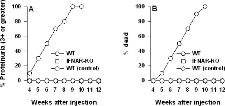
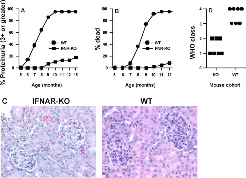
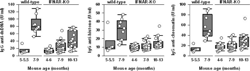
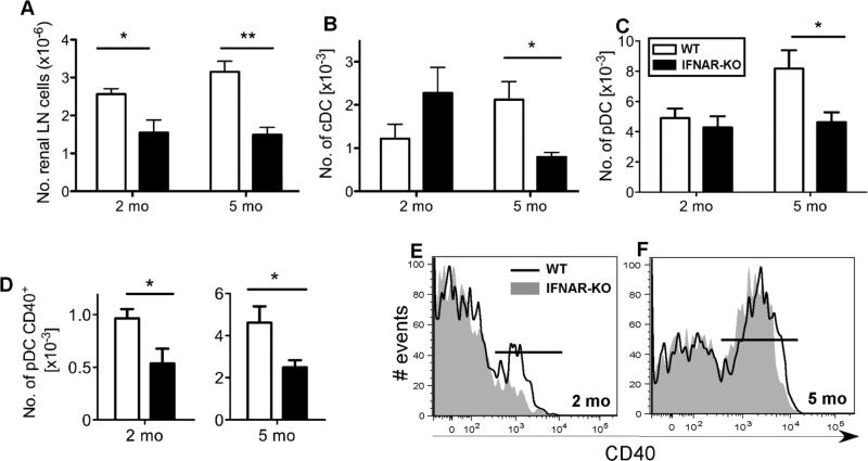
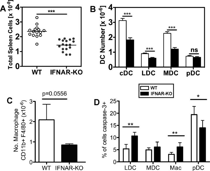
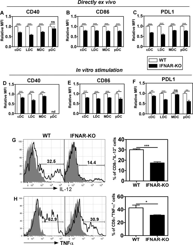
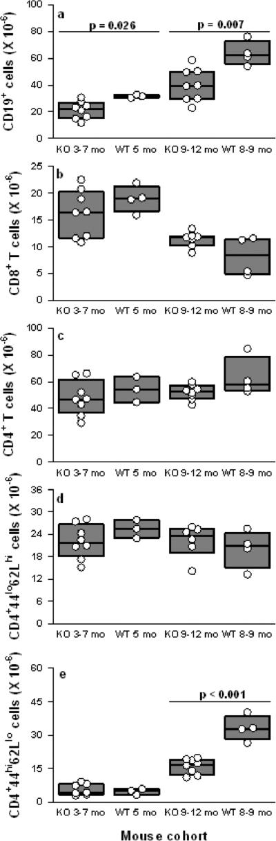
Type I IFNs are potent regulators of innate and adaptive immunity and are implicated in the pathogenesis of systemic lupus erythematosus. Here we report that clinical and pathological lupus nephritis and serum anti-nuclear Ab levels are greatly attenuated in New Zealand Mixed (NZM) 2328 mice deficient in type I IFN receptors (IFNAR). To determine whether the inflammatory environment in NZM 2328 mice leads to IFNAR-regulated changes in dendritic cells (DC), the number, activation, and function of DC subsets were compared in 2- and 5-mo-old (clinically healthy) female NZM and NZM-IFNAR(-/-) mice. Numbers of activated CD40(high) plasmacytoid DC (pDC) were significantly increased in renal lymph nodes of 2-mo-old NZM but not NZM-IFNAR(-/-) mice, suggesting an early IFNAR-dependent expansion and activation of pDC at disease sites. Relative to NZM spleens, NZM-IFNAR(-/-) spleens in 5-mo-old mice were significantly decreased in size and contained reduced numbers of conventional DC subsets, but not pDC. Splenic and renal lymph node NZM-IFNAR(-/-) DC analyzed directly ex vivo expressed significantly less CD40, CD86, and PDL1 than did NZM DC. Upon activation with synthetic TLR9 ligands in vitro, splenic NZM-IFNAR(-/-) DC produced less IL-12p40/70 and TNF-alpha than did NZM DC. The limited IFNAR(-/-) DC response to endogenous activating stimuli correlated with reduced numbers of splenic activated memory CD4(+) T cells and CD19(+) B cells in older mice. Thus, IFNAR signaling significantly increases DC numbers, acquisition of Ag presentation competence, and proinflammatory function before onset of clinically apparent lupus disease.


Figures

Fig. 1
Fig. 1. In vivo adenovirus-mediated delivery of mIFNα fails to induce proteinuria and death in NZM 2328 mice deficient in IFNAR
8-9 week old WT NZM 2328 female mice (n=16) and IFNAR deficient (IFNAR-KO) NZM 2328 mice (n=13) were injected with IFNα-producing adenovirus as described in M&M. A third group of WT female mice (n=7) labeled “control” were injected with an adenovirus lacking the IFNα construct. Data are presented as the percent cumulative prevalence of severe (+3, >300mg/dl) proteinuria and as percent cumulative mortality at the ages shown.
Fig. 2
Fig. 2. IFNAR deficiency protects NZM 2328 mice from proteinuria, kidney pathology, and mortality
Shown are cumulative (A) proteinuria and (B) mortality of NZM 2328 WT and IFNAR-deficient (KO) female mice with age. A total of 21-24 mice for each group were monitored up to 12 mo of age. Data are presented as the percent cumulative prevalence of severe (+3, >300mg/dl) proteinuria and as percent cumulative mortality at the ages shown. (C) A representative H&E stained kidney section (×400) from an 11 mo old IFNAR-deficient female mouse (Left) showing mild glomerular infiltration, is compared to an 8.5 mo old WT NZM 2328 female mouse (Right) showing marked glomerular hypercellularity. (D) Quantification of GN was done using the WHO scoring system (class 1 to class 4). IFNAR-KO mice at 10-12 months of age were compared to WT mice at 7-9 months of age. Please note that WT of older age were not available because these mice die by this age.
Fig. 3
Fig. 3. Delayed production of IgG autoantibodies in IFNAR-deficient mice
Comparison of serum IgG anti-dsDNA, anti-histone, and anti-chromatin Ab levels in WT NZM 2328 mice and IFNAR-deficient female mice at the indicated ages. The composite results are plotted as box plots, with median (line inside box), 25th and 75th percentiles (box boundaries) and 10th and 90th percentiles (bars extending from boxes) indicated. All three types of autoantibodies are significantly higher in 7-9 month old WT mice compared to IFNAR-/- mice at 7-9 or 10-13 months of age with p<0.001.
Fig. 4
Fig. 4. Two and five month old NZM 2328 mice show an IFNAR-dependent increase in activated pDC in renal lymph nodes
(A) IFNAR deficiency decreases the total number of rLN cells. rLN were digested with collagenase and total viable cell numbers determined by hemocytometer counts. (B) Numbers of rLN cDC are reduced in 5 month old IFNAR-KO mice. (C) Numbers of rLN pDC (gated as shown in Fig. S1) are reduced in 5 month old IFNAR-KO mice. (D) Numbers of activated CD40hi pDC are reduced in 2 and 5 month old IFNAR-KO mice. For panels A-D, shown are the mean + SEM values for WT (open bar) and IFNAR-KO (closed bar) cells, n=5; *p<0.05, **p<0.01. (E) The percentage of rLN pDC expressing high levels of CD40 is shown for representative 2 month old WT (18%, black line) and IFNAR-KO (10%, shaded gray) mice. (F) The percentage of rLN pDC expressing high levels of CD40 is shown for representative 5 month old WT (57%, black line) and IFNAR-KO (56%, shaded gray) mice.
Fig. 5
Fig. 5. Reduction of numbers of total splenocytes, splenic macrophages, and splenic cDC, but not pDC, in 5 month old IFNAR-/- mice
(A) IFNAR deficiency reduces the splenomegaly characteristic of NZM 2328 mice. Spleens were digested with collagenase, red cells lysed and total viable cell numbers determined by hemocytometer counts. Shown are the values for individual 5 month old mice with the mean value indicated by the bar. The mean + SEM for WT spleens was 2.36 + 0.104 ×10⁸, n=15 and for IFNAR-KO spleens was 1.43 + 0.089 ×10⁸, n=17, ***p<0.0001. (B) Numbers of splenic cDC but not pDC were reduced in IFNARKO mice. Splenic DC subsets were identified using multi-parameter flow cytometry according to the gating scheme shown in Fig. S1. Shown are the mean + SEM numbers of each DC subset, n=15-17; ***p<0.0001; ns, not significant. (C) Numbers of splenic macrophages tend to be reduced in IFNAR-KO mice. Shown are the mean + SEM, n=5; p=0.0556. (D) IFNAR deficiency alters the percentages of myeloid cells that express activated caspase-3. Shown are the mean + SEM of each cell subset, n=5; *p<0.05, **p<0.01.
Fig. 6
Fig. 6. IFNAR-/- DC are less activated in vivo and show a reduced capacity for activation upon TLR9 stimulation in vitro
(A-C) Splenic DC from IFNAR-KO mice were less activated in vivo. Splenic DC subsets, as gated in Fig. S1, were analyzed directly ex vivo for surface expression of (A) CD40 (B) CD86 and (C) PDL1. (D-F) Splenic DC from IFNAR-KO mice showed less upregulation of costimulatory molecules after in vitro activation by TLR9 ligands. Splenic DC were stimulated for 16 hr in vitro with CpG-B ODN (5 μg/ml) and DC subsets analyzed for expression of (D) CD40, (E) CD86 and (F) PDL1. For panels A-F, shown are mean + SEM of relative MFI values of each costimulatory molecule on the indicated splenic DC subsets in WT (open bar) or IFNAR-KO (closed bar) mice. The data were compiled from 3-4 independent experiments, each with groups of 4-5 mice (20 weeks of age), using a statistical normalization method. Raw data were log-transformed to achieve normality and/or to satisfy the equal variance test. *p<0.05; **p<0.01; ***p<0.001; nd, not determined. (G) IFNAR-KO LDC produced less IL-12p40/70 than WT LDC. Splenocytes were activated in vitro for 18 hr with CpG-B ODN (5 μg/ml), with brefeldin A added for the last 10 hr, prior to detection of IL-12+ LDC by surface and intracellular staining. Shown are representative histograms of IL-12p40/70 expression (black line) as compared to unstimulated cells (shaded histogram), and the mean + SEM values for WT and IFNAR-KO DC, n=4-5; ***p<0.0001. Data are representative of 3 independent experiments. (H) IFNAR-KO LDC produced less TNFα than WT LDC. Splenocytes were activated in vitro for 6 hr with CpG-B ODN (5 μg/ml), in the presence of brefeldin A, prior to detection of TNFα+ LDC. Shown are representative histograms of TNFα expression (black line) as compared to unstimulated cells (shaded histogram), and the mean + SEM values for WT and IFNAR-KO DC, n=4; *p=0.039. Data are representative of 2 independent experiments.
Fig. 7
Fig. 7. IFNAR deficiency prevents expansion of splenic CD19+ B cells and activated memory CD4+ T cells
For B and T lymphocyte subsets, numbers shown are millions of cells per spleen of WT or IFNAR-KO mice at the indicated ages. Older (10-12 month) WT mice were not available because the majority had died by 9-10 months of age. The composite results are plotted as box plots. The lines inside the boxes indicate the medians; the outer borders of the boxes indicate 25th and 75th percentiles; the bars extending from the boxes indicate the 10th and 90th percentiles. Flow cytometry was used to identify (A) CD19+ B lymphocytes; (B) CD8+ T cells; (C) CD4+ T cells; (D) CD4+CD44loCD62Lhigh naive T cells and (E) CD4+CD44highCD62Llow/neg activated memory T cells. Significant differences between IFNAR-deficient and WT mice are indicated in the plots.

References

    1. Theofilopoulos AN, Baccala R, Beutler B, Kono DH. Type I interferons (alpha/beta) in immunity and autoimmunity. Annu Rev Immunol. 2005;23:307–336. - PubMed
    1. Baccala R, Hoebe K, Kono DH, Beutler B, Theofilopoulos AN. TLR-dependent and TLR-independent pathways of type I interferon induction in systemic autoimmunity. Nat Med. 2007;13:543–551. - PubMed
    1. Honda K, Takaoka A, Taniguchi T. Type I interferon gene induction by the interferon regulatory factor family of transcription factors. Immunity. 2006;25:349–360. - PubMed
    1. Kadowaki N, Antonenko S, Lau JY, Liu YJ. Natural interferon alpha/beta-producing cells link innate and adaptive immunity. J Exp Med. 2000;192:219–226. - PMC - PubMed
    1. Liu YJ, Kanzler H, Soumelis V, Gilliet M. Dendritic cell lineage, plasticity and cross-regulation. Nat Immunol. 2001;2:585–589. - PubMed

Publication types

MeSH terms