. 2009 Dec;20(23):4997-5006.
doi: 10.1091/mbc.e09-04-0304. Epub 2009 Oct 7.

Tight functional coupling of kinesin-1A and dynein motors in the bidirectional transport of neurofilaments

Affiliations

Tight functional coupling of kinesin-1A and dynein motors in the bidirectional transport of neurofilaments

Atsuko Uchida et al. Mol Biol Cell. 2009 Dec.

Abstract

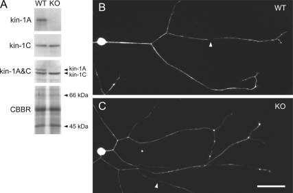
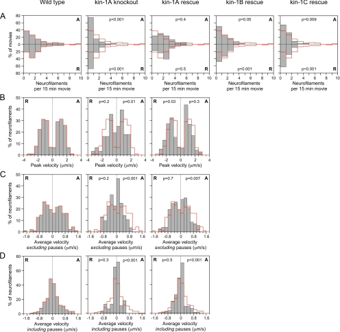
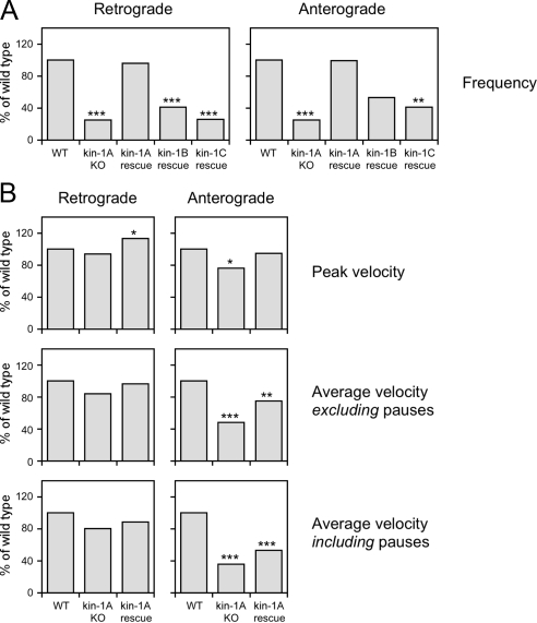
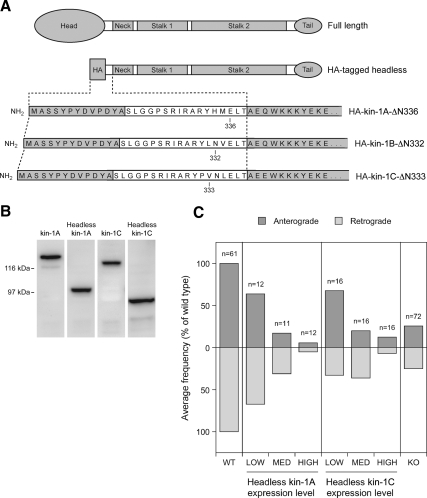
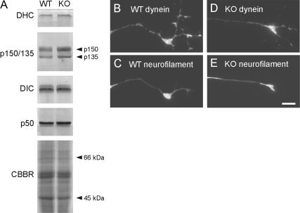
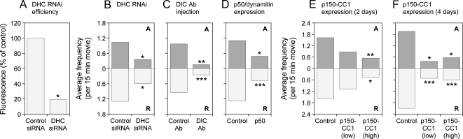
We have tested the hypothesis that kinesin-1A (formerly KIF5A) is an anterograde motor for axonal neurofilaments. In cultured sympathetic neurons from kinesin-1A knockout mice, we observed a 75% reduction in the frequency of both anterograde and retrograde neurofilament movement. This transport defect could be rescued by kinesin-1A, and with successively decreasing efficacy by kinesin-1B and kinesin-1C. In wild-type neurons, headless mutants of kinesin-1A and kinesin-1C inhibited both anterograde and retrograde movement in a dominant-negative manner. Because dynein is thought to be the retrograde motor for axonal neurofilaments, we investigated the effect of dynein inhibition on anterograde and retrograde neurofilament transport. Disruption of dynein function by using RNA interference, dominant-negative approaches, or a function-blocking antibody also inhibited both anterograde and retrograde neurofilament movement. These data suggest that kinesin-1A is the principal but not exclusive anterograde motor for neurofilaments in these neurons, that there may be some functional redundancy among the kinesin-1 isoforms with respect to neurofilament transport, and that the activities of the anterograde and retrograde neurofilament motors are tightly coordinated.


Figures

Figure 1.
Figure 1.
Neurons from kinesin-1A knockout mice extend axons containing neurofilaments. (A) Western blotting of total brain protein from wild-type (WT) and kinesin-1A knockout (KO) littermates, confirming the absence of kinesin-1A. Kinesin-1A and kinesin-1C were detected using affinity purified antisera. Mixing these two antisera (kinesin-1A&C) confirmed the absence of kinesin-1A because kinesin-1A migrates more slowly than kinesin-1C by SDS-PAGE. To demonstrate equivalent protein content of the two samples, we show a portion of a gel stained with Coomassie Brilliant Blue R (CBBR). (B) Cultured SCG neurons from wild type and kinesin-1A knockout mouse littermates after 12 h in culture, immunostained with a polyclonal antibody against neurofilament protein L. Neurofilaments extended throughout the axons of both wild-type and knockout neurons, occasionally interrupted by short gaps (for example, see white arrowheads). Bar, 50 μm.
Figure 2.
Figure 2.
Reduced frequency and velocity of neurofilament movement in neurons from kinesin-1A knockout mice. Neurofilament transport was analyzed by tracking the movement of neurofilaments through gaps in the axonal neurofilament array in wild-type neurons (196 filaments in 61 movies), kinesin-1A knockout neurons (59 filaments in 72 movies), and in kinesin-1A knockout neurons expressing either kinesin-1A (149 filaments in 46 movies; kin-1A rescue), kinesin-1B (84 filaments observed in 54 movies; kin-1B rescue), or kinesin-1C (51 filaments in 46 movies; kin-1C rescue). (A) Frequency of neurofilament movement plotted as neurofilaments per 15-min movie (all movies were exactly 15 min in length). The anterograde (A) and retrograde (R) frequencies are represented by the top and bottom bars, respectively. (B–D) Peak and average velocities of neurofilament movement. In each graph, the anterograde velocities (A) are shown on the right and the retrograde velocities (R) on the left. The wild-type distributions (see red line) are superimposed on each graph to aid comparison of the data. The p values represent the probability that the wild-type and experimental distributions arose from the same populations (Mann–Whitney test). See Supplemental Tables S1 and S2 for a more detailed summary of these data.
Figure 3.
Figure 3.
Summary of kinetic data. (A) Bar graphs showing the mean frequency of neurofilament movement in wild-type neurons (WT); in kinesin-1A knockout neurons (kin-1A KO); and in kinesin-1A knockout neurons expressing kinesin-1A (kin-1A rescue), kinesin-1B (kin-1B rescue), or kinesin-1C (kin-1C rescue), each normalized to the mean in the wild type. (B) Bar graphs showing the mean peak velocity and mean average velocity including or excluding pauses for wild-type neurons (WT), kinesin-1A knockout neurons (kin-1A KO), and for kinesin-1A knockout neurons expressing kinesin-1A (kin-1A rescue), each normalized to the mean in the wild type. The asterisks denote probability that the wild-type and experimental distributions arose from the same populations (Mann–Whitney test: ***p < 0.005, **p < 0.01, and *p < 0.05). See Supplemental Tables S1 and S2 for a more detailed summary of these data.
Figure 4.
Figure 4.
Headless kinesin-1A and kinesin-1C inhibit movement in both directions. (A) Schematic illustration of the structure of the headless kinesin-1A, B, and C constructs. Each construct was created by replacing the N-terminal head domain with an HA epitope tag, but retaining the entire tail, stalk, and neck domains and a portion of the neck linker (Kozielski et al., 1997). The sequence MASSYPYDVPDYA is the HA tag and the sequence SLGGPSRIRARY in the linker comes from the multiple cloning site of the HA vector. Kinesin-1A is truncated at amino acid 336, kinesin-1B at amino acid 332, and kinesin-1C at amino acid 333. (B) Western blot of extracts from cultured SW13 cells expressing full-length and headless kinesin-1A and C constructs, stained with mAb H2. The apparent molecular masses are ∼131 and 98 kDa for full-length and HA-tagged headless KIFA, respectively, and ∼122 and 92 kDa for full-length and HA-tagged headless KIFC, respectively. (C) Cultured SCG neurons from wild-type mice were cotransfected by nuclear injection with GFP-NFM and either HA-tagged headless kinesin-1A or HA-tagged headless kinesin-1C. Neurofilament transport was analyzed by time-lapse fluorescence imaging of gaps. To correlate the extent of neurofilament movement with the level of headless construct expression, we fixed the cultures after live-cell imaging and processed them for immunostaining using antibodies against NFM and the HA tag (see Materials and Methods). The bar graphs show the correlation between the average frequency of neurofilament movement (normalized to the frequency in the wild type) and the expression level of the headless construct. The anterograde and retrograde frequencies are represented by the top and bottom bars, respectively. The average frequency of movement in the wild type (WT) and kinesin-1A knockout (KO) in the absence of headless construct are included for comparison.
Figure 5.
Figure 5.
Dynein and dynactin levels are normal in brains and in axons of cultured neurons from kinesin-1A knockout mice. (A) Western blot of total brain proteins from wild-type (WT) and kinesin-1A knockout (KO) mice obtained with antibodies specific for dynein heavy chain (DHC), dynein intermediate chain (DIC), and the p150/135 and p50 subunits of dynactin. To demonstrate equivalent protein content of the two samples, we show a portion of a gel stained with Coomassie Brilliant Blue R (CBBR). (B–E) Immunostaining of wild-type and knockout SCG neurons using antibodies specific for dynein intermediate chain (B and D) and neurofilament protein M (C and E). Bar, 5 μm.
Figure 6.
Figure 6.
Disruption of dynein/dynactin inhibits both anterograde and retrograde neurofilament transport. Effect of disruption of dynein/dynactin on the average frequency of neurofilament movement in SCG neurons (A–D) and cortical neurons (E and F) from wild-type mice. Data for each condition are the average of at least 20 15-min movies gathered from at least three different experiments. The anterograde (A) and retrograde (R) frequencies are represented by the top and bottom bars, respectively. (A) Efficiency of RNAi knockdown after 7 d culture (6 d after siRNA injection), determined by immunostaining with an antibody to dynein heavy chain (see Materials and Methods). Control siRNA: cells injected with scrambled siRNA (26 movies). DHC siRNA: cells injected with a pool of three siRNAs targeting dynein heavy chain (21 movies). (B) Effect of dynein heavy chain knockdown on neurofilament movement 6 d after siRNA injection (7 d in culture). Control siRNA: cells injected with scrambled siRNA (23 movies). DHC siRNA: cells injected with a pool of three siRNAs targeting dynein heavy chain (20 movies). (C) Effect of function-blocking dynein intermediate chain antibody IC74.1 on neurofilament movement 2–6 h after antibody injection (5 d in culture). Control antibody (Ab): cells injected with control IgG (38 movies). DIC Ab: cells injected with antibody IC74.1 (20 movies). (D) Effect of p50/dynamitin overexpression on neurofilament movement 3 d after transfection (5 d in culture). Control: untreated cells (59 movies). p50: cells transfected with p50/dynamitin construct (44 movies). (E) Effect of p150-CC1 overexpression on neurofilament movement 2 d after transfection (2 d in culture). Control: untreated cells (26 movies). p150-CC1 (low): cells transfected with 25 μg/ml p150-CC1 construct (23 movies). p150-CC1 (high): cells transfected with 75 μg/ml p150-CC1 construct (20 movies). (F) Effect of p150-CC1 overexpression on neurofilament movement 4 d after transfection (4 d in culture). Control: untreated cells (20 movies). p150-CC1 (low): cells transfected with 25 μg/ml p150-CC1 construct (21 movies). p150-CC1 (high): cells transfected with 75 μg/ml p150-CC1 construct (21 movies). The data in A were compared to the control with a t test. The data in B–F were compared to the control with the Mann–Whitney test. p values: ***p < 0.005, **p < 0.01, and *p < 0.05. See Supplemental Tables S3 and S4 for a more detailed summary of these data.

References

    1. Ahmad F. J., He Y., Myers K. A., Hasaka T. P., Francis F., Black M. M., Baas P. W. Effects of dynactin disruption and dynein depletion on axonal microtubules. Traffic. 2006;7:524–537. - PubMed
    1. Bloom G. S., Wagner M. C., Pfister K. K., Brady S. T. Native structure and physical properties of bovine brain kinesin and identification of the ATP-binding subunit polypeptide. Biochemistry. 1988;27:3409–3416. - PubMed
    1. Brady S. T., Pfister K. K., Bloom G. S. A monoclonal antibody against kinesin inhibits both anterograde and retrograde fast axonal transport in squid axoplasm. Proc. Natl. Acad. Sci. USA. 1990;87:1061–1065. - PMC - PubMed
    1. Brown A. Slow axonal transport: stop and go traffic in the axon. Nat. Rev. Mol. Cell. Biol. 2000;1:153–156. - PubMed
    1. Brown A. Axonal transport of membranous and nonmembranous cargoes: a unified perspective. J. Cell Biol. 2003;160:817–821. - PMC - PubMed

Publication types

MeSH terms

LinkOut - more resources