. 2010 Feb;24(2):627-36.
doi: 10.1096/fj.09-142281. Epub 2009 Oct 7.

Control of oxidative phosphorylation by vitamin A illuminates a fundamental role in mitochondrial energy homoeostasis

Affiliations

Control of oxidative phosphorylation by vitamin A illuminates a fundamental role in mitochondrial energy homoeostasis

Rebeca Acin-Perez et al. FASEB J. 2010 Feb.

Abstract

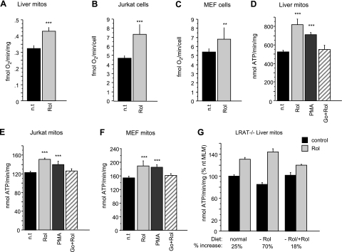
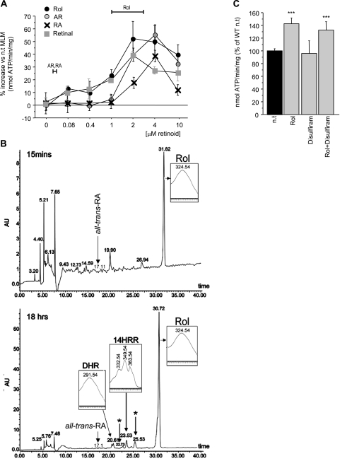
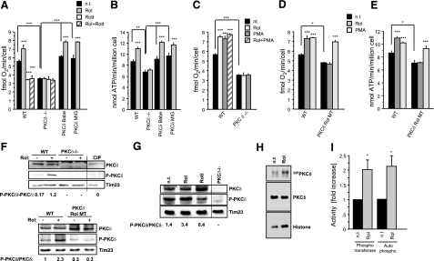
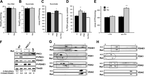
The physiology of two metabolites of vitamin A is understood in substantial detail: retinaldehyde functions as the universal chromophore in the vertebrate and invertebrate eye; retinoic acid regulates a set of vertebrate transcription factors, the retinoic acid receptor superfamily. The third member of this retinoid triumvirate is retinol. While functioning as the precursor of retinaldehyde and retinoic acid, a growing body of evidence suggests a far more fundamental role for retinol in signal transduction. Here we show that retinol is essential for the metabolic fitness of mitochondria. When cells were deprived of retinol, respiration and ATP synthesis defaulted to basal levels. They recovered to significantly higher energy output as soon as retinol was restored to physiological concentration, without the need for metabolic conversion to other retinoids. Retinol emerged as an essential cofactor of protein kinase Cdelta (PKCdelta), without which this enzyme failed to be activated in mitochondria. Furthermore, retinol needed to physically bind PKCdelta, because mutation of the retinol binding site rendered PKCdelta unresponsive to Rol, while retaining responsiveness to phorbol ester. The PKCdelta/retinol complex signaled the pyruvate dehydrogenase complex for enhanced flux of pyruvate into the Krebs cycle. The baseline response was reduced in vitamin A-deficient lecithin:retinol acyl transferase-knockout mice, but this was corrected within 3 h by intraperitoneal injection of vitamin A; this suggests that vitamin A is physiologically important. These results illuminate a hitherto unsuspected role of vitamin A in mitochondrial bioenergetics of mammals, acting as a nutritional sensor. As such, retinol is of fundamental importance for energy homeostasis. The data provide a mechanistic explanation to the nearly 100-yr-old question of why vitamin A deficiency causes so many pathologies that are independent of retinoic acid action.


Figures

Figure 1.
Figure 1.
Retinol modulates respiration. A) Pyruvate/malate-dependent oxygen consumption in retinol-treated (Rol; 2 μM, 10 min) mouse liver mitochondria (MLM) is increased by 30% compared to untreated mitochondria (n=6) (n.t., not treated). B, C) Respiration in intact retinol-treated (4 μM, 2 h) Jurkat (B) or MEF (C) cells is increased by 60 and 30%, respectively, compared to nontreated cells (Jurkat cells, n=14; MEF cells, n=8). D–F) Pyruvate/malate-driven ATP synthase activity is increased in isolated MLM (D; n=12) or mitochondria of Jurkat cells (E; n=6) or MEF cells (F; n=6). Mitochondria were incubated for 10 min with 2 μM retinol, phorbol-myristoyl-acetate (PMA, 2 μg/ml), or the combination of Go6976 (5 μM) and retinol (2 μM). Retinol increased ATP synthesis in the 3 systems by 55% (MLM), 25% (Jurkat mitochondria), and 23% (MEF mitochondria). Retinol effect was mimicked by PMA, but was neutralized by the PKC inhibitor, Go6976. G) Comparison of retinol-mediated up-regulation of ATP synthase activity of MLM isolated from mice with differing vitamin A nutritional status. LRAT−/− mice were maintained on vitamin A-deficient or -sufficient diets. One group of vitamin A-deficient mice was reconstituted by IP injection of vitamin A 3 h before harvesting liver. MLM were tested for ATP synthetic rate before and after incubation with 2 μM retinol in organello for 10 min. Average relative increases (%) were plotted. Retinol stimulated ATP synthase activity more profoundly in LRAT−/− mitochondria of vitamin A-deprived mice (70%) than those of deficient (25%), reconstituted LRAT−/−, or wild-type mice (not shown). **P < 0.001; ***P < 0.0001.
Figure 2.
Figure 2.
Capacity of natural retinoids to stimulate ATP synthase activity and their bioavailability in cells. A) Dose-response characteristics of retinol (Rol), retinal, retinoic acid (RA), and anhydroretinol (AR) are similar. However, only retinol bioavailability matches the dose optimum needed for synthase activation (horizontal bars). B) Retinoid profile, determined by high-pressure liquid chromatography. No retinol metabolites were found in MEFs incubated for 15 min with 5 μM retinol (top panel), whereas after 18 h, MEFs contained 13,14-dihydroxyretinol (DHR), 14-hydroxy-retro-retinol, and 2 unidentified retinoids (asterisk), but no discernible retinoic acid. Positive identification was by UV spectra (inserts) and mass spectrometry (not shown). C) Disulfiram does not block retinol-mediated ATP synthase stimulation.
Figure 3.
Figure 3.
PKCδ is a target for retinol action in mitochondria. A) Cellular respiration in wild-type (WT) MEFs (n=9), PKCδ−/−MEFs (n=9), or PKCδ−/− MEFs, where wild-type PKCδ was reintroduced by retroviral vectors, PKCδ rBabe (n=6), and PKCδ rMIG (n=6). Retinol (Rol) stimulated respiration in wild-type but not in PKCδ−/− MEFs. This stimulation was blocked by the PKCδ-specific inhibitor rottlerin (Rott). Retroviral reintroduction of full-length PKCδ in PKCδ−/− MEFs rescued responsiveness to retinol. B) Pyruvate/malate-driven ATP synthase activity in wild-type (n=10), PKCδ−/− (n=10), or PKCδ−/− reconstituted (n=6/cell line). Retinol stimulated ATP synthesis in wild-type but not in PKCδ−/− MEFs. Reintroduction of PKCδ in PKCδ−/− MEFs rescued the retinol response. C) Up-regulation of oxygen consumption was mimicked by PMA and was dependent on PKCδ. PMA and retinol effects were not additive. D, E) PKCδ requires intact retinol binding sites. PKCδ−/− MEFs reconstituted with full-length mutated PKCδ gene were unable to bind retinol (PKCδ Rol MT) and failed to restore responsiveness to retinol, as determined by respiration (D) and ATP synthase activity (E), respectively. F) Western blotting of wild-type MEF cell homogenates revealed that PKCδ was phosphorylated at Thr505 to higher degree when retinol was present, compared to its absence (top panel). MEFs expressing the retinol nonbinding mutant PKCδ did not yield appreciably phosphorylated PKCδ (bottom panel). Absence of immunoreactivity to PKCδ−/− MEF extracts indicated specificity of phosphoThr505-antibody reaction. Tim 23 was used as gel-loading control. Densitometry values of the intensity ratios P- PKCδ/ PKCδ are displayed beneath blots (n=3). G) PKCδ activation by retinol in isolated mitochondria. Stimulation of isolated wild-type mitochondria with retinol resulted in increased PKCδ phosphorylation. Rottlerin inhibited retinol-mediated PKCδ phosphorylation. PKCδ−/− MEFs are shown as specificity control. H) PKCδ kinase activity is stimulated by retinol. Wild-type MEFs were retinol deprived in serum-free medium. Aliquots were either left untreated or supplemented with 2 μM retinol. Mitochondria were isolated and lysed, and PKCδ activity was determined by immunoprecipitation/kinase assay . Phosphotransferase activity was revealed by PKC autophosphorylation (autoradiograph of top panel) and histone heterophosphorylation (autoradiograph of bottom panel). Images were analyzed by densitometry and normalized on amounts of PKC immunoprecipitated (Western blot of middle panel). I) Results from H presented as fold increases over basal PKC levels. One of 3 representative experiments is shown. *P < 0.01; **P < 0.001; ***P < 0.0001.
Figure 4.
Figure 4.
Retinol regulates PDH activity through PKCδ activation. A–C) Oxygen consumption driven by glutamate/malate (A) or succinate (n=6) (B), or ATP synthase activity driven by succinate (n=12) (C) in MLM were unchanged on retinol stimulation (2 μM, 10 min). D) PDH activity measured spectrophotometrically in MLM (n=9). Retinol stimulation increased PDH activity by 30%. Retinol effect was mimicked by PMA, but inhibited by Go6976. E) Enhanced pyruvate/malate-dependent ATP synthesis in MLM treated with retinol compared to untreated mitochondria was not observed when phosphatase inhibitors (Phi) were present, but was evident when Phi were absent (n=6). F) Western blots of MEF mitochondria revealed decreased PDHE1 phosphorylation as opposed to increased PKCδ phosphorylation at Thr505 when retinol was present in mitochondria of wild-type cells. Alkaline phosphatase treatment (CIP) of wild-type (WT) mitochondria was used to ascertain the specificity of the P-PKCδ and P-PDHE1 antibodies. Tim 23 was used as loading control. Intensity of bands was quantified by densitometry; ratio of phosphorylated/total products is displayed beneath blots (n=3). G, H) Two-dimensional gel electrophoresis revealed retinol-induced changes of phosphoproteome. Mitochondrial proteins were separated by IEF followed by SDS-PAGE of wild-type (G) or PKCδ−/− (H) MEF cells. PDHE1 was less phosphorylated in wild-type retinol-treated than untreated (n.t.) mitochondria (G, top blot). PDHE1 phosphorylation state remained unchanged in PKCδ−/− mitochondria, when treated with retinol (H, top blot). PDK1 lost phosphate groups in both wild-type and PKCδ−/− mitochondria on retinol exposure (G, H; second blot from top). PDK2 reduced its phosphorylation state in retinol-treated wild-type mitochondria, compared to untreated wild-type mitochondria and retinol-treated or untreated PKCδ−/− mitochondria (G, H; third from top). VDAC was used as a reference marker to position the blots. (See also Supplemental Fig. 2.) Blots are representative of 3 independent experiments. *P < 0.01; **P < 0.001; ***P < 0.0001.

References

    1. McCollum E V, Davis M. The necessity of certain lipins in the diet during growth. J Biol Chem. 1913;15:167–175. - PubMed
    1. Osborne T B, Mendel L B. The relation of growth to the chemical constitutents of the diet. J Biol Chem. 1913;145:311–326.
    1. Green S, Chambon P. Nuclear receptors enhance our understanding of transcription regulation. Trends Genet. 1988;4:309–314. - PubMed
    1. Evans R M, Hollenberg S M. Zinc-fingers: guilt by association. Cell. 1988;52:1–3. - PubMed
    1. Wald G. Molcular basis of visual excitation. Science. 1968;162:230–239. - PubMed

Publication types

MeSH terms