. 2010 Mar;16(3):867-78.
doi: 10.1089/ten.TEA.2009.0288.

Age-related changes in material behavior of porcine mitral and aortic valves and correlation to matrix composition

Affiliations

Age-related changes in material behavior of porcine mitral and aortic valves and correlation to matrix composition

Elizabeth H Stephens et al. Tissue Eng Part A. 2010 Mar.

Abstract

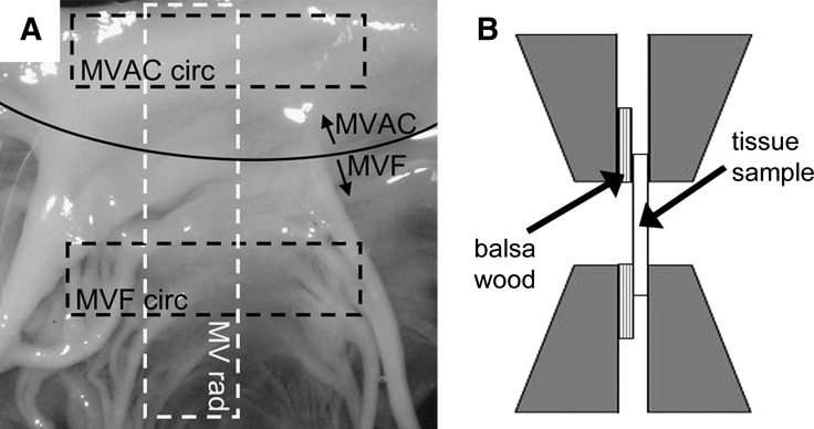
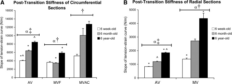
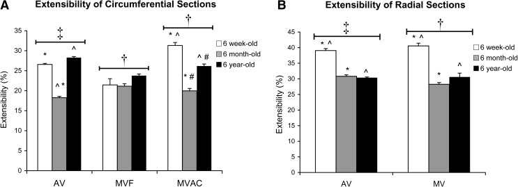
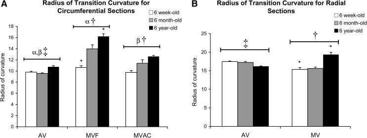
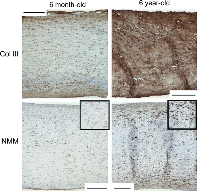
Recent studies showing significant changes in valvular matrix composition with age offer design criteria for age-specific tissue-engineered heart valves. However, knowledge regarding aging-related changes in valvular material properties is limited. Therefore, 6-week, 6-month, and 6-year-old porcine aortic valves (AV) and mitral valves (MV) were subjected to uniaxial tensile testing. In addition to standard material parameters, the radius of transition curvature (RTC) was measured to assess the acuteness of the transition region of the tension-strain curve. Radially, the MV had greater stiffness and a smaller RTC compared with the AV. Circumferentially, the center of the MV anterior leaflet (MVAC) had the highest stiffness (MVAC > AV > MV free edge [MVF]), greater stress relaxation (MVAC > MVF/AV), lowest extensibility (MVAC < AV < MVF), and smaller RTC compared with MVF (AV < MVAC < MVF). AV and MV radial strips had a larger RTC compared with circumferential strips. Aging elevated stiffness for MV and AV radial and circumferential strips, elevated stress relaxation in AV and MVF circumferential strips, and increased RTC for MV radial and MVF circumferential strips. In conclusion, there are significant age-related differences in the material properties of heart valves, which parallel differences in tissue composition and structure, likely impact valve function, and highlight the need for age-specific design goals for tissue-engineered heart valves.


Conflict of interest statement

No competing financial interests exist.

Figures

FIG. 1.
FIG. 1.
(A) Orientation of tissue strips cut from the mitral valve (MV) (indicated by dashed boxes). Curved solid line indicates the border between the center (MVAC) and free edge (MVF) of the mitral value (MV) anterior leaflet. Rad, radial; circ, circumferential. (B) Diagram illustrating the placement of glued balsa/valve tissue sample construct within grips of material testing system.
FIG. 2.
FIG. 2.
(A) Thickness of circumferential valve strips with age. Results for radial strips were comparable (data not shown). (B) Thickness of circumferential valve strips with age normalized to heart size (maximum circumference of heart). *,^Significantly different between ages within a given valve region. α,βValve regions significantly different for a given direction (radial or circumferential).
FIG. 3.
FIG. 3.
Slope of post-transition region of tension–strain curves for (A) circumferential strips and (B) radial strips. *,^Significantly different between ages within a given valve region by post hoc ANOVA comparing all ages. αAV and MV significantly different for a given direction (radial or circumferential). AV radial significantly different from AV circumferential; MVAC circumferential, MVF circumferential, and MV radial strips each significantly different from one another. AV, aortic valve; ANOVA, analysis of variance.
FIG. 4.
FIG. 4.
Slope of the pre-transition region of tension–strain curves for (A) circumferential strips and (B) radial strips. *,^Significantly different between ages within a given valve region by post hoc ANOVA comparing all ages. α,βAV and MV significantly different for a given direction (radial or circumferential).
FIG. 5.
FIG. 5.
Total percentage of relaxation (SR) for (A) circumferential strips and (B) radial strips. *,^Significantly different between ages within a given valve region by post hoc ANOVA comparing all ages. αValve regions significantly different for a given direction (radial or circumferential). AV radial significantly different from AV circumferential. MVAC circumferential, MVF circumferential, and MV radial strips each significantly different from one another.
FIG. 6.
FIG. 6.
Extensibility for (A) circumferential strips and (B) radial strips. *,^,#Significantly different between ages within a given valve region by post hoc ANOVA comparing all ages. AV radial significantly different from AV circumferential. MV radial strips are significantly different from both MVAC and MVF circumferential strips.
FIG. 7.
FIG. 7.
Radius of transition curvature: (A) circumferential strips and (B) radial strips. *Significantly different between ages within a given valve region by post hoc ANOVA comparing all ages. α,βAV and MV significantly different for a given direction (radial or circumferential). AV radial significantly different from AV circumferential. MVAC circumferential, MVF circumferential, and MV radial strips each significantly different from one another.
FIG. 8.
FIG. 8.
Representative Movat-stained tissue sample sections for 6-week-old and 6-year-old radial strips. In Movat pentachrome-stained tissue, yellow = aligned collagen, black = elastic fibers, and green/blue = proteoglycans (PGs)/glycosaminoglycans (GAGs). To facilitate comparison between AV and MV, valves were all oriented with the fibrosa at the bottom of the image and sized to show the complete leaflet thickness allowing visualization of all leaflet layers. Because of differences in leaflet thickness (see Fig. 2), magnifications differ between images. Scale bars equal 200 μm. Note that the MV radial annulus is almost exclusively fibrosa (corresponding to the MVAC), whereas AV radial annulus shows a considerably smaller proportion of the leaflet thickness composed of fibrosa. Also of note is the marbling of PG/GAG with collagen in the fibrosa of 6-year-old MV radial annular strips (see close-up view in inset) and MVAC circumferential strips compared with 6-week-old strips. This marbling may, in part, account for the increase in SR seen with age (see Discussion section). Color images available online at www.liebertonline.com/ten.
FIG. 9.
FIG. 9.
Representative Movat-stained tissue sample sections for 6-week-old, 6-month-old, and 6-year-old circumferential strips. In Movat pentachrome-stained tissue, yellow = aligned collagen, black = elastic fibers, and green/blue = PGs/GAGs. To facilitate comparison between AV and MV, valves were all oriented with the fibrosa at the bottom of the image. Images in column 1 show high magnification of 6-week-old valves (all images in column 1 are the same magnification). Images in columns 2–4 are lower magnification to demonstrate the differences in leaflet thickness (all images in columns 2–4 are the same magnification). Scale bars equal 200 μm. Compared with MVF, MVAC circumferential sections contain much higher proportion of fibrosa. For all valve regions an increase in collagen was noted throughout the valve layers with age, but most particularly in the fibrosa and ventricularis. Color images available online at www.liebertonline.com/ten.
FIG. 10.
FIG. 10.
MVAC circumferential sections showing increased Col III and NMM in 6-year-old sections compared with 6-month-old sections. Similar differences were noted for the staining of Col III for other valve regions between the 6-year-old aged sections and the younger ages. Scale bar equals 200 μm. Insets in NMM images are 1.5 × higher magnification for improved visualization of cells. Col III, collagen type III; NMM, nonmuscle myosin heavy chain-IIB. Color images available online at www.liebertonline.com/ten.

References

    1. Keller F. Leutert G. [Age dependence of collagen structures of the human heart] Z Gerontol. 1994;27:186. - PubMed
    1. Angrist A. Aging heart valves and a unitary pathological hypothesis for sclerosis. J Gerontol. 1964;19:135. - PubMed
    1. Aikawa E. Whittaker P. Farber M. Mendelson K. Padera R.F. Aikawa M. Schoen F.J. Human semilunar cardiac valve remodeling by activated cells from fetus to adult: implications for postnatal adaptation, pathology, and tissue engineering. Circulation. 2006;113:1344. - PubMed
    1. Bashey R.I. Torii S. Angrist A. Age-related collagen and elastin content of human heart valves. J Gerontol. 1967;9:203. - PubMed
    1. Sell S. Scully R.E. Aging changes in the aortic and mitral valves. Histologic and histochemical studies, with observations on the pathogenesis of calcific aortic stenosis and calcification of the mitral annulus. Am J Pathol. 1965;46:345. - PMC - PubMed

Substances