. 2009 Oct 8:6:26.
doi: 10.1186/1743-8977-6-26.

Diesel exhaust particles modulate the tight junction protein occludin in lung cells in vitro

Affiliations

Diesel exhaust particles modulate the tight junction protein occludin in lung cells in vitro

Andrea D Lehmann et al. Part Fibre Toxicol. .

Abstract

Background: Using an in vitro triple cell co-culture model consisting of human epithelial cells (16HBE14o-), monocyte-derived macrophages and dendritic cells, it was recently demonstrated that macrophages and dendritic cells create a transepithelial network between the epithelial cells to capture antigens without disrupting the epithelial tightness. The expression of the different tight junction proteins in macrophages and dendritic cells, and the formation of tight junction-like structures with epithelial cells has been demonstrated. Immunofluorescent methods combined with laser scanning microscopy and quantitative real-time polymerase chain reaction were used to investigate if exposure to diesel exhaust particles (DEP) (0.5, 5, 50, 125 mug/ml), for 24 h, can modulate the expression of the tight junction mRNA/protein of occludin, in all three cell types.

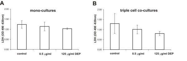
Results: Only the highest dose of DEP (125 mug/ml) seemed to reduce the occludin mRNA in the cells of the defence system however not in epithelial cells, although the occludin arrangement in the latter cell type was disrupted. The transepithelial electrical resistance was reduced in epithelial cell mono-cultures but not in the triple cell co-cultures, following exposure to high DEP concentration. Cytotoxicity was not found, in either epithelial mono-cultures nor in triple cell co-cultures, after exposure to the different DEP concentrations.

Conclusion: We concluded that high concentrations of DEP (125 mug/ml) can modulate the tight junction occludin mRNA in the cells of the defence system and that those cells play an important role maintaining the epithelial integrity following exposure to particulate antigens in lung cells.


Figures

Figure 1
Figure 1
Internalized DEP in an epithelial cell. TEM images of 16HBE14o- cells in the triple cell co-culture model showing internalized DEP. Cells were exposed to 125 μg/ml DEP (A) and 0.5 μg/ml DEP (B) for 24 h and then processed for TEM. A detailed view from A and B (white square) is shown in A'and B'.
Figure 2
Figure 2
TJ between EC and between EC and MDM. TEM images showing untreated cells in the triple cell co-culture model in A. The tight junctions (TJ) the adherens junctions (AJ), as well as the desomosomes (D) between two neighbouring EC are shown in the detailed view in A1. TJ-like structures (white arrows, B1 and B2) between a MDM and two neighbouring EC in the triple cell co-culture model is shown in overview in B. B1 and B2 are detailed views. The morphology and as well as the internalized gold NP (black arrow in B2) prove that the described cell is a MDM.
Figure 3
Figure 3
Laser scanning micrographs of occludin in EC, MDM and MDDC. The occludin arrangement in 16HBE14o-, MDM and MDDC mono-cultures visualized with LSM. Control 16HBE14o- cells (A) and DEP exposed 16HBE14o- cells (B) were labelled for F-actin (turquoise) and occludin (red). For F-actin no qualitative difference was found between control and treated cells. However, the iso-surface presentation of occludin in control cells (A') showed a more structured TJ occludin belt compared with DEP treated cells (B") which shows a predominant localization of occludin in the cytoplasm. Occludin was visualized in mono-cultures of MDM (D, control; C, DEP treatment) and MDDC (E, control; F, DEP treatment). Cells were stained for F-actin (in MDM white, in MDDC black and occludin (shown in red). MDM and MDDC expressed the TJ protein occludin. After exposure to DEP for 24 h occludin signals were reduced (for MDM in D' and MDDC in F'). These are representative images and for all pictures the same settings were used.
Figure 4
Figure 4
mRNA expression levels of occludin in EC, MDM and MDDC. Quantification of occludin mRNA expression in MDM, MDDC, and EC mono-cultures by qRT-PCR. Threshold of occludin was set by the threshold of the housekeeping gene β-actin in each sample (ΔCT). Expression levels of occludin are illustrated by the β-actin normalized concentrations (2-ΔCT × 15000). cDNA derived from MDM (grey columns), MDDC (black columns) and 16HBE14o- (white columns) control cells and cells treated with DEP (0.5 μg/ml, 5 μg/ml, 50 μg/ml, and 125 μg/ml). Values are presented as means ± SD; * p ≤ 0.05. All experiments were independently performed between 4-7 times.
Figure 5
Figure 5
Epithelial integrity of EC mono- and triple cell co-cultures. TEER in16HBE14o- mono-cultures (A) and triple cell co-cultures (B) before (white bars) and after (black bars) exposure to high dose of DEP (125 μg/ml) or normal medium (control) for 24 h. TEER in mono-cultures was significantly lower after DEP treatment than before addition of DEP (A, *). Data are presented as means ± standard deviation (SD); * p ≤ 0.05. All experiments were independently done in triplicate.
Figure 6
Figure 6
Cytotoxicity of EC mono- and triple cell co-cultures. LDH (a marker for membrane permeability) levels were determined. The values did not change significantly after DEP exposure in epithelial mono-cultures (A) and triple cell co-cultures (B). Data are presented as mean ± SD of 3 experiments.
Figure 7
Figure 7
TNFα release in EC mono- and triple cell co-cultures. TNFα release in mono-cultures (A) and triple cell co-cultures (B) upon exposure to DEP for 24 h. TNFα concentrations were expressed as mean ± SD of 4-5 experiments. DEP treated EC mono-cultures and the triple cell co-cultures showed no difference in TNFα concentration in comparison to the respective untreated cells. No differences between mono-cultures and triple cell co-cultures concerning TNFα levels were found. LPS stimulated cultures were used for the positive control.
Figure 8
Figure 8
Opening of the TJ in EC mono- and triple cell co-cultures. TEER values after EDTA treatment (grey lines) over one hour in mono-cultures (A) and in triple cell co-cultures (B). Untreated controls are presented in black.

Similar articles

Cited by

References

    1. Pope CA, III, Thun MJ, Namboodiri MM, Dockery DW, Evans JS, Speizer FE, et al. Particulate air pollution as a predictor of mortality in a prospective study of U.S. adults. Am J Respir Crit Care Med. 1995;151:669–674. - PubMed
    1. Peters A, Wichmann HE, Tuch T, Heinrich J, Heyder J. Respiratory effects are associated with the number of ultrafine particles. Am J Respir Crit Care Med. 1997;155:1376–1383. - PubMed
    1. Schulz H, Harder V, Ibald-Mulli A, Khandoga A, Koenig W, Krombach F, et al. Cardiovascular effects of fine and ultrafine particles. J Aerosol Med. 2005;18:1–22. doi: 10.1089/jam.2005.18.1. - DOI - PubMed
    1. Ruckerl R, Phipps RP, Schneider A, Frampton M, Cyrys J, Oberdorster G, et al. Ultrafine particles and platelet activation in patients with coronary heart disease--results from a prospective panel study. Part Fibre Toxicol. 2007;4:1. doi: 10.1186/1743-8977-4-1. - DOI - PMC - PubMed
    1. Bayram H, Devalia JL, Sapsford RJ, Ohtoshi T, Miyabara Y, Sagai M, et al. The effect of diesel exhaust particles on cell function and release of inflammatory mediators from human bronchial epithelial cells in vitro. Am J Respir Cell Mol Biol. 1998;18:441–448. - PubMed