. 2009 Dec;37(12):1411-22.
doi: 10.1016/j.exphem.2009.09.009. Epub 2009 Oct 6.

Polycythemia vera erythroid precursors exhibit increased proliferation and apoptosis resistance associated with abnormal RAS and PI3K pathway activation

Affiliations

Polycythemia vera erythroid precursors exhibit increased proliferation and apoptosis resistance associated with abnormal RAS and PI3K pathway activation

Jacob P Laubach et al. Exp Hematol. 2009 Dec.

Abstract

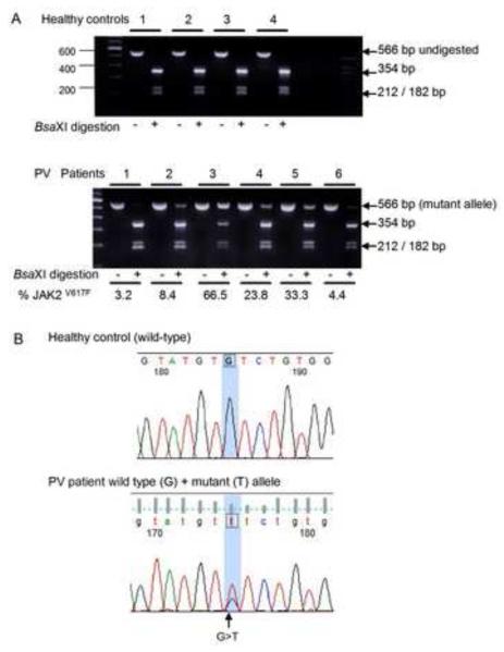
Objective: Polycythemia vera (PV) is characterized by erythrocytosis associated with the presence of the activating JAK2(V617F) mutation in a variable proportion of hematopoietic cells. JAK2(V617F) is detected in other myeloproliferative neoplasms, does not appear to be the PV-initiating event, and its specific role in deregulated erythropoiesis in PV is incompletely understood. We investigated the pathogenesis of PV to characterize abnormal proliferation and apoptosis responses and aberrant oncogenic pathway activation in primary PV erythroid precursors.

Materials and methods: Peripheral blood CD34(+) cells isolated from PV patients and healthy controls were grown in liquid culture to expand a population of primary erythroblasts for experiments designed to analyze cellular proliferation, apoptosis, JAK2(V617F) mutation status, cytokine-dependent protein phosphorylation and gene expression profiling using Affymetrix microarrays.

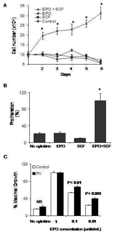
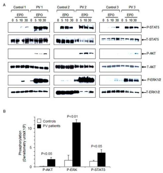
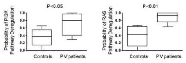
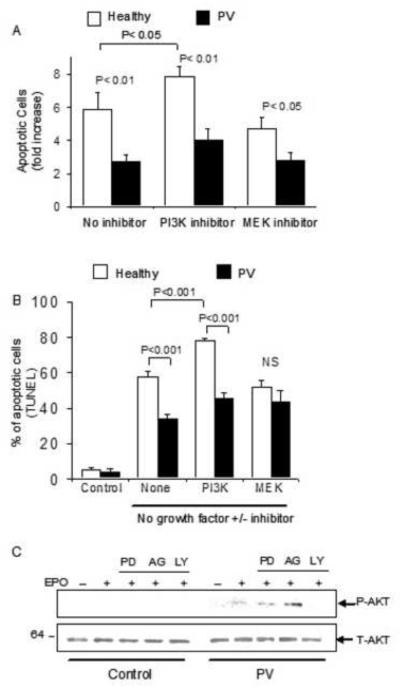
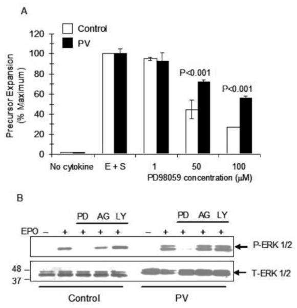
Results: The survival and proliferation of PV erythroblasts were growth factor-dependent under strict serum-free conditions requiring both erythropoietin (EPO) and stem cell factor. PV erythroblasts exhibited EPO hypersensitivity and enhanced cellular proliferation associated with increased EPO-mediated extracellular signal-regulated kinases 1 and 2 phosphorylation. EPO-induced AKT phosphorylation was observed in PV but not normal erythroblasts, an effect associated with apoptosis resistance in PV erythroblasts. Analysis of gene expression and oncogenic pathway activation signatures revealed increased RAS (p<0.01) and phosphoinositide-3 kinase (p<0.05) pathway activation in PV erythroblasts.

Conclusion: Deregulated erythropoiesis in PV involves EPO hypersensitivity and apoptosis resistance of erythroid precursor cells associated with abnormally increased activation of RAS-ERK and phosphoinositide-3 kinase-AKT pathways. These data suggest that investigation of the mechanisms of abnormal RAS and phosphoinositide-3 kinase pathway activation in erythroblasts may contribute to our understanding of the molecular pathogenesis of PV.


Figures

Figure 1
Figure 1. Detection of JAK2V617F mutation in primary erythroid precursors
Total RNA isolated from primary erythroblasts of healthy controls and PV patients was reverse transcribed followed by PCR amplification for JAK2 yielding a 566 bp product. (A) Detection of JAK2V617F by restriction enzyme BsaXI digestion. The wild-type allele is digested into 354, 212, and 182 bp fragments whereas the mutant allele, detected only in PV patients, remains undigested. The allele-burden of JAK2V617F in PV patients was quantified by densitometry and expressed as percentage. (B) Sequence traces illustrating wild-type sequence (a healthy control) and confirmation of mixed G>T sequences in a representative patient with PV. The presence of the mutant JAK2V617F allele was confirmed by direct sequencing in all patient samples.
Figure 2
Figure 2. Proliferation of PV erythroid precursors in response to cytokines under serum-free conditions
(A) Growth curves of PV erythroblasts plated in serum-free medium in the presence of EPO, SCF or both growth factors in a representative experiment. Cells were counted daily in a hemocytometer using trypan blue exclusion technique and total cell counts were plotted. The data represent mean ± SEM values of triplicate cultures. *P<0.001 (B) Short-term expansion of erythroblasts in serum-free cultures (four days) in response to cooperative effect of EPO and SCF determined by MTT assay. Control group contained no cytokines. Data represent mean ± SEM values (n=6 replicates in each group). *P<0.001. (C) EPO hypersensitivity of PV erythroblasts in serum-free conditions containing decreasing concentrations of EPO ranging as indicated with assessment of proliferation in an MTT assay. Data are represented as % of maximal growth. NS: not significant.
Figure 3
Figure 3. Phosphorylation of STAT5, AKT and ERK1/2, in primary erythroblasts in response to EPO
(A) Proerythroblasts were washed free of serum and growth factors, incubated in serum-free starvation medium and then either left unstimulated (0) or stimulated with EPO (10 units/mL) for the indicated time period (5, 10, 30 minutes). Whole cell lysates were analyzed by immunoblotting. Increased EPO-induced phosphorylation of STAT5, AKT and ERK1/2 are noted. Comparable protein loading in each lane and protein integrity were demonstrated by stripping the blots and hybridizing to an antibody that detects total protein levels. (B) Quantification and comparison of levels of phosphorylated STAT5, AKT, and ERK1/2 in PV versus control erythroblasts stimulated with EPO at 10 minutes. Densitometry measurements were performed and represented as mean ± SEM from three PV patients and three healthy controls.
Figure 4
Figure 4. Analysis of RAS and PI3-Kinase pathway activation signatures
Gene expression signatures of oncogenic signaling pathways to characterize the activation status of RAS and PI3-kinase pathways were applied to data from patients with PV and healthy controls. The probability of PI3-kinase (panels on left) and RAS pathway (panels on right) deregulation in PV erythroblasts are significantly increased compared to normal control erythroblasts.
Figure 5
Figure 5. PV erythroid precursors exhibit reduced apoptosis following growth factor withdrawal
Proerythroblasts from PV patients and healthy individuals were induced to undergo apoptosis by growth factor (EPO, SCF, IGF-I) withdrawal in the presence or absence of kinase inhibitors for 16 hours. Apoptosis was assessed using (A) Annexin V assays, expressed as fold-increase in apoptotic cells compared to erythroblasts cultured in complete erythroblast medium supplemented with EPO, SCF and IGF-I as control, and (B) TUNEL assays. Control cultures containing complete medium with growth factor exhibit low basal apoptotic cell percentage that is significantly increased following growth factor withdrawal for 16 hours. (C) EPO-induced AKT phosphorylation was observed only in PV erythroblasts and not in healthy control cells. AKT phosphorylation is inhibited by treatment with PI3-kinase inhibitor LY but not after treatment with kinase inhibitors targeting MEK (PD) or JAK2 (AG).
Figure 6
Figure 6. Decreased sensitivity of PV erythroblasts to the growth-inhibitory effect of MEK kinase inhibitor compared to normal erythroblasts
(A) PV or healthy control erythroblasts were placed in serum-free medium for short-term expansion (four days) in the presence of EPO and SCF (E+S) and the indicated concentrations of MEK inhibitor PD98059. Proliferation was measured by MTT assay. Control group contained no growth factors. E+S group contained DMSO vehicle. (B) EPO-induced ERK phosphorylation was inhibited by treatment of erythroblasts with MEK inhibitor PD98059 (PD) but not after treatment with kinase inhibitors targeting PI3-kinase (LY) or JAK2 (AG).

Similar articles

Cited by

References

    1. Prchal JF, Axelrad AA. Letter: Bone-marrow responses in polycythemia vera. N Engl J Med. 1974;290:1382. - PubMed
    1. Eaves CJ, Eaves AC. Erythropoietin (Ep) dose-response curves for three classes of erythroid progenitors in normal human marrow and in patients with polycythemia vera. Blood. 1978;52:1196–1210. - PubMed
    1. Casadevall N, Vainchenker W, Lacombe C, et al. Erythroid progenitors in polycythemia vera: demonstration of their hypersensitivity to erythropoietin using serum free cultures. Blood. 1982;59:447–451. - PubMed
    1. Means RT, Jr., Krantz SB, Sawyer ST, Gilbert HS. Erythropoietin receptors in polycythemia vera. J Clin Invest. 1989;84:1340–1344. - PMC - PubMed
    1. Hess G, Rose P, Gamm H, Papadileris S, Huber C, Seliger B. Molecular analysis of the erythropoietin receptor system in patients with polycythaemia vera. Br J Haematol. 1994;88:794–802. - PubMed

Publication types

MeSH terms