Review
. 2009:278:309-53.
doi: 10.1016/S1937-6448(09)78007-4.

Polarity proteins and cell-cell interactions in the testis

Affiliations
Review

Polarity proteins and cell-cell interactions in the testis

Elissa W P Wong et al. Int Rev Cell Mol Biol. 2009.

Abstract

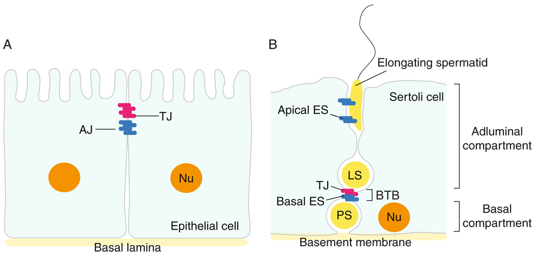
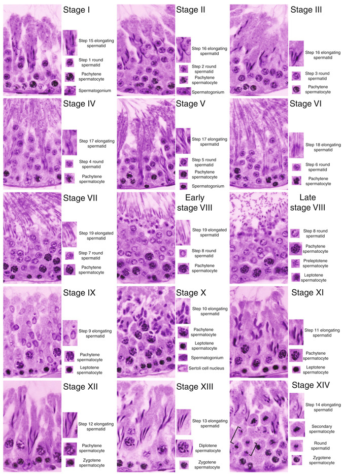
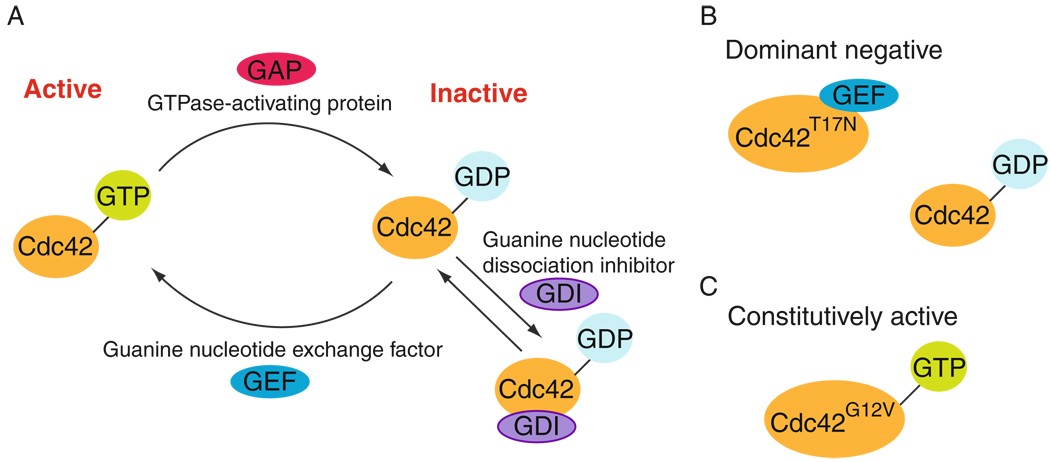
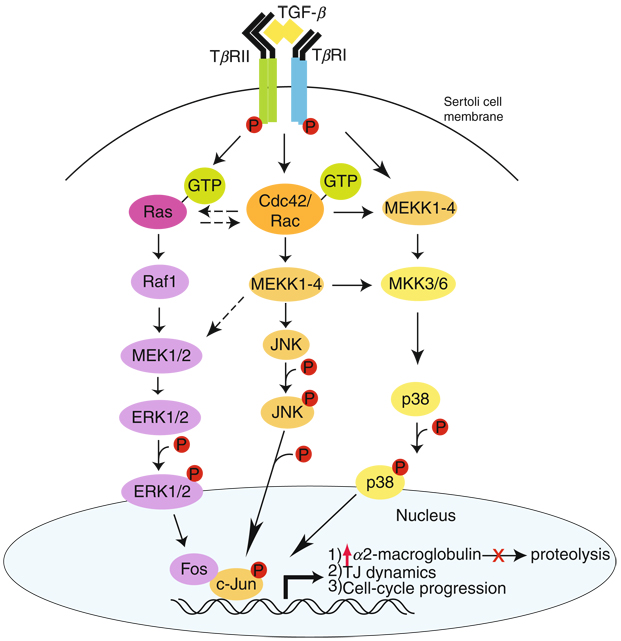
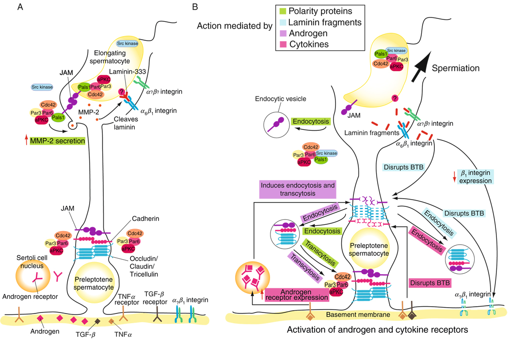
In mammalian testes, extensive junction restructuring takes place in the seminiferous epithelium at the Sertoli-Sertoli and Sertoli-germ cell interface to facilitate the different cellular events of spermatogenesis, such as mitosis, meiosis, spermiogenesis, and spermiation. Recent studies in the field have shown that Rho GTPases and polarity proteins play significant roles in the events of cell-cell interactions. Furthermore, Rho GTPases, such as Cdc42, are working in concert with polarity proteins in regulating cell polarization and cell adhesion at both the blood-testis barrier (BTB) and apical ectoplasmic specialization (apical ES) in the testis of adult rats. In this chapter, we briefly summarize recent findings on the latest status of research and development regarding Cdc42 and polarity proteins and how they affect cell-cell interactions in the testis and other epithelia. More importantly, we provide a new model in which how Cdc42 and components of the polarity protein complexes work in concert with laminin fragments, cytokines, and testosterone to regulate the events of cell-cell interactions in the seminiferous epithelium via a local autocrine-based regulatory loop known as the apical ES-BTB-basement membrane axis. This new functional axis coordinates various cellular events during different stages of the seminiferous epithelium cycle of spermatogenesis.


Figures

Figure 7.1
Figure 7.1
Differences in the spatial arrangement of tight junction (TJ) and adherens junction (AJ) in epithelia versus the seminiferous epithelium in adult mammalian testes. (A) In most epithelia, TJ is restricted to the apical region of adjacent epithelial cells, underneath of which lies AJ, a cell–cell actin-based anchoring junction type. Furthest away from TJ is the basal lamina, a form of extracellular matrix (ECM). In other blood–tissue barrier, the TJ alone confers the barrier function, such as in the blood–brain barrier and the blood–retina barrier. (B) In mammalian testes, such as rats, the blood–testis barrier (BTB) is closest to the basement membrane (a modified form of ECM) instead of the apical region of the Sertoli cell. Also, the BTB is composed of coexisting TJ and basal ES (an atypical AJ type in the testis). The apical ES, however, is restricted to the Sertoli cell–elongating spermatid interface. The presence of the BTB also segregates the seminiferous epithelium into the basal and adluminal compartments.
Figure 7.2
Figure 7.2
Different stages of the seminiferous epithelial cycle of spermatogenesis in adult rat testes. Stages I through XIV of the epithelial cycle in adult rat testes are shown. The typical germ cells at different stages of their development that are found in specific stage of the epithelial cycle are shown on the right panel of the corresponding micrographs. For instance, both meiosis I and II take place in Stage XIV in rat testes, so that secondary spermatocytes following meiosis I and round spermatids (step 1) frommeiosis II following telephase II (see square brackets) are clearly visible.
Figure 7.3
Figure 7.3
The cycling of Cdc42 GTPase between GTP- (active) and GDP-bound (inactive) state. (A) The activation of Cdc42 GTPase involves the exchange of GDP for GTP via phosphorylation, which is stimulated by GEF (guanine nucleotide exchange factor), leading to an increase in affinity of activated Cdc42 for its effector to stimulate downstream signaling functions. Activated Cdc42 GTPase can be inactivated by its binding with GTPase-activating protein (GAP), leading to dephosphorylation, and a shutdown of the signaling function. The release of GDP from the Cdc42 GTPase is blocked by guanine nucleotide dissociation inhibitor (GDI). The GDI-bound Cdc42 GTPase is sequestered in the cytosol. (B) This illustrates a dominant-negative form of Cdc42 in which the threonine (Thr, T) in residue 17 from the N-terminus is mutated to asparagine (Asn, N), which allows binding of GEF but not effectors. Thus, GEF is sequestered by the dominant-negative form and this prevents endogenous Cdc42 from activating by GEF (Bollag and McCormick, 1991; Heasman and Ridley, 2008). (C) This is the constitutively active form of Cdc42 wherein the glycine (Gly, G) in residue 12 from the N-terminus is mutated to valine (Val, V), which is defective in GTPase activity, thus it cannot be dephosphorylated (i.e., inactivated) but remains phosphorylated (activated). Other common constitutively active mutants include mutation at residue 18 from phenylalanine (Phe, F) to leucine (Leu, L) and mutation at residue 61 from glutamine (Gln, Q) to leucine (Leu, L) (Bollag and McCormick, 1991; Heasman and Ridley, 2008).
Figure 7.4
Figure 7.4
The TGF-β-mediated signaling function in epithelia including the seminiferous epithelium of adult rat testes. Based on recent studies in the field, TGF-βs are known to regulate different cellular functions in the testis following activation of their receptors via ligand-receptor binding, such as for cell cycle progression in germ cells, TJ dynamics, and α2-macroglobulin production, via ERK, p38 MAP or JNK (black arrows). This schematic diagram illustrates the significance of Cdc42 in TGF-βs-mediated signaling function since this GTPase regulates not only the JNK signaling pathway downstream, also the ERK1/2 and p38 MAPK pathways via its cross talk with Ras and MEK1/2 (hatched arrows) or its direct effects on MEKK1–4 and MKK3/6 (black arrows).
Figure 7.5
Figure 7.5
The three highly conserved polarity protein complexes: the partitioning defective (PAR), Crumbs (CRB) and Scribble complexes, that are found in multiple epithelia including the seminiferous epithelium in rat testes. Many components of these proteins are also found in germ cells in the seminiferous epithelium. Interaction between Par6 and Pals1 provides cross talk between the CRB and Par complexes (black arrows). aPKC is a crucial component in the polarity protein complexes which provides cross talk between the three conserved polarity complexes. Phosphorylation of Lgl by aPKC maintains the Scribble complex at the basolateral domain (solid line bars).
Figure 7.6
Figure 7.6
Schematic drawing illustrating the involvement of cytokines, testosterone, biologically active fragments of laminin chains, hemidesmosome, and polarity proteins in regulating spermiation and BTB restructuring during the seminiferous epithelial cycle of spermatogenesis. This schematic drawing was prepared based on recent findings in the field as detailed in the text. (A) In this panel, the known protein complexes at the apical ES, namely the JAM-C-based protein complex and the α6-β1-integrin/ laminin-333-based protein complex are shown. The cell adhesion at the BTB is conferred by the JAM-A-based, cadherin-based, and the occludin-, claudin-, and tricellulin- based protein complexes. Just prior to spermiation, the polarity protein complex, Cdc42/Par3/Par6/Pals1/aPKC, remains associated with JAM-C. The presence of the polarity complex is likely involved in targeting and activating MMP-2 at the apical ES, which apparently is being used to cleave the laminin chains to generate the biologically active fragments. (B) During spermiation at the apical ES, Src kinase was shown to associate more tightly with Par6 and Pals1, causing the dissociation of the Par-based polarity complex from the JAM-C-based protein complex. JAM-C may also be internalized via endocytosis due to the absence of Par6 at the apical ES at spermiation, further destabilizing the JAM-C-based adhesion and facilitating spermiation at stage VIII of the epithelial cycle. Laminin fragments generated at the apical ES site were shown to perturb the BTB integrity directly or indirectly, acting as autocrine factors, via their effects on a yet-to-be identified integrin receptor at the BTB, and β1-integrin at the hemidesmosome. At the BTB, the biologically active laminin fragments apparently accelerate endocytic-vesicle-mediated endocytosis of integral membrane proteins, destabilizing the “old” TJ-fibrils above a primary preleptotene spermatocyte in transit at the BTB at stage VIII of the epithelial cycle. Cytokines, such as TGF-β2 and TGF-β3, are also likely to be involved in “destabilizing” the “old” BTB by accelerating endocytosis of BTB integral membrane proteins above the primary spermatocytes in transit. However, the combined effects of testosterone and TNFα-induced androgen receptor expression may accelerate the production (e.g., de novo synthesis of occludin, claudins, and JAMs) and assembly of “new” TJ-fibrils behind a primary spermatocyte in transit, and by transcytosing junction proteins from the “old” barrier to new site behind the spermatocyte. The processes of protein endocytosis and recycling, and perhaps transcytosis, are facilitated by polarity protein components, such as Par3, Par6, and 14-3-3. The polarity protein complex may serve as initial spatial cue to direct endocytosed proteins for forming “new” barrier behind the primary spermatocyte. Through the combined and concerted efforts of cytokines, testosterone, and biologically active laminin fragments, and with the participation of polarity proteins via their actions on protein endocytosis, recycling, and transcytosis, “new” TJ-fibrils can be formed behind a primary spermatocyte in transit prior to the dissolution of the “old” TJ-fibrils. Thus, the BTB is being restructured to facilitate the transit of spermatocytes while the immunological barrier can be maintained during spermatogenesis.

References

    1. Adams AE, Johnson DI, Longnecker RM, Sloat BF, Pringle JR. CDC42 and CDC43, two additional genes involved in budding and the establishment of cell polarity in the yeast Saccharomyces cerevisiae. J. Cell Biol. 1990;111:131–142. - PMC - PubMed
    1. Aitken A. 14-3-3 proteins: a historic overview. Semin. Cancer Biol. 2006;16:162–172. - PubMed
    1. Al-Sadi R, Boivin M, Ma T. Mechanism of cytokine modulation of epithelial tight junction barrier. Front Biosci. 2009;14:2765–2778. - PMC - PubMed
    1. Andras IE, Pu H, Deli MA, Nath A, Hennig B, Toborek M. HIV-1 Tat protein alters tight junction protein expression and distribution in cultured brain endothelial cells. J. Neurosci. Res. 2003;74:255–265. - PubMed
    1. Ang AL, Taguchi T, Francis S, Folsch H, Murrells LJ, Pypaert M, et al. Recycling endosomes can serve as intermediates during transport from the Golgi to the plasma membrane of MDCK cells. J. Cell Biol. 2004;167:531–543. - PMC - PubMed

Publication types

LinkOut - more resources