Functional interchangeability of rod and cone transducin alpha-subunits
- PMID: 19815523
- PMCID: PMC2758286
- DOI: 10.1073/pnas.0901382106
Functional interchangeability of rod and cone transducin alpha-subunits
Abstract
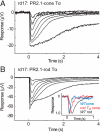
Rod and cone photoreceptors use similar but distinct sets of phototransduction proteins to achieve different functional properties, suitable for their role as dim and bright light receptors, respectively. For example, rod and cone visual pigments couple to distinct variants of the heterotrimeric G protein transducin. However, the role of the structural differences between rod and cone transducin alpha subunits (Talpha) in determining the functional differences between rods and cones is unknown. To address this question, we studied the translocation and signaling properties of rod Talpha expressed in cones and cone Talpha expressed in rods in three mouse strains: rod Talpha knockout, cone Talpha GNAT2(cpfl3) mutant, and rod and cone Talpha double mutant rd17 mouse. Surprisingly, although the rod/cone Talpha are only 79% identical, exogenously expressed rod or cone Talpha localized and translocated identically to endogenous Talpha in each photoreceptor type. Moreover, exogenously expressed rod or cone Talpha rescued electroretinogram responses (ERGs) in mice lacking functional cone or rod Talpha, respectively. Ex vivo transretinal ERG and single-cell recordings from rd17 retinas treated with rod or cone Talpha showed comparable rod sensitivity and response kinetics. These results demonstrate that cone Talpha forms a functional heterotrimeric G protein complex in rods and that rod and cone Talpha couple equally well to the rod phototransduction cascade. Thus, rod and cone transducin alpha-subunits are functionally interchangeable and their signaling properties do not contribute to the intrinsic light sensitivity differences between rods and cones. Additionally, the technology used here could be adapted for any such homologue swap desired.
Conflict of interest statement
Conflict of interest statement: W.W.H. and the University of Florida have a financial interest in the use of AAV therapies and own equity in a company (AGTC Inc.) that might, in the future, commercialize some aspects of this work.
Figures


Similar articles
-
Replacing the rod with the cone transducin subunit decreases sensitivity and accelerates response decay.J Physiol. 2010 Sep 1;588(Pt 17):3231-41. doi: 10.1113/jphysiol.2010.191221. Epub 2010 Jul 5. J Physiol. 2010. PMID: 20603337 Free PMC article.
-
Diminished Cone Sensitivity in cpfl3 Mice Is Caused by Defective Transducin Signaling.Invest Ophthalmol Vis Sci. 2020 Apr 9;61(4):26. doi: 10.1167/iovs.61.4.26. Invest Ophthalmol Vis Sci. 2020. PMID: 32315379 Free PMC article.
-
Transretinal ERG recordings from mouse retina: rod and cone photoresponses.J Vis Exp. 2012 Mar 14;(61):3424. doi: 10.3791/3424. J Vis Exp. 2012. PMID: 22453300 Free PMC article.
-
Tuning outer segment Ca2+ homeostasis to phototransduction in rods and cones.Adv Exp Med Biol. 2002;514:179-203. doi: 10.1007/978-1-4615-0121-3_11. Adv Exp Med Biol. 2002. PMID: 12596922 Review.
-
Photoreceptor physiology and evolution: cellular and molecular basis of rod and cone phototransduction.J Physiol. 2022 Nov;600(21):4585-4601. doi: 10.1113/JP282058. Epub 2022 Apr 28. J Physiol. 2022. PMID: 35412676 Free PMC article. Review.
Cited by
-
Activation and quenching of the phototransduction cascade in retinal cones as inferred from electrophysiology and mathematical modeling.Mol Vis. 2015 Mar 7;21:244-63. eCollection 2015. Mol Vis. 2015. PMID: 25866462 Free PMC article.
-
Exchange of Cone for Rod Phosphodiesterase 6 Catalytic Subunits in Rod Photoreceptors Mimics in Part Features of Light Adaptation.J Neurosci. 2015 Jun 17;35(24):9225-35. doi: 10.1523/JNEUROSCI.3563-14.2015. J Neurosci. 2015. PMID: 26085644 Free PMC article.
-
Replacing the rod with the cone transducin subunit decreases sensitivity and accelerates response decay.J Physiol. 2010 Sep 1;588(Pt 17):3231-41. doi: 10.1113/jphysiol.2010.191221. Epub 2010 Jul 5. J Physiol. 2010. PMID: 20603337 Free PMC article.
-
Mouse Models of Inherited Retinal Degeneration with Photoreceptor Cell Loss.Cells. 2020 Apr 10;9(4):931. doi: 10.3390/cells9040931. Cells. 2020. PMID: 32290105 Free PMC article. Review.
-
A kinetic analysis of mouse rod and cone photoreceptor responses.J Physiol. 2020 Sep;598(17):3747-3763. doi: 10.1113/JP279524. Epub 2020 Jul 14. J Physiol. 2020. PMID: 32557629 Free PMC article.
References
-
- Korenbrot JI, Rebrik TI. Tuning outer segment Ca2+ homeostasis to phototransduction in rods and cones. Adv Exp Med Biol. 2002;514:179–203. - PubMed
-
- Kawamura S, Tachibanaki S. Rod and cone photoreceptors: Molecular basis of the difference in their physiology. Comp Biochem Physiol A Mol Integr Physiol. 2008;150:369–377. - PubMed
-
- Chang B, et al. Cone photoreceptor function loss-3, a novel mouse model of achromatopsia due to a mutation in Gnat2. Invest Ophthalmol Vis Sci. 2006;47:5017–5021. - PubMed
-
- Burns ME, Arshavsky VY. Beyond counting photons: Trials and trends in vertebrate visual transduction. Neuron. 2005;48:387–401. - PubMed
Publication types
MeSH terms
Substances
Grants and funding
LinkOut - more resources
Full Text Sources
Molecular Biology Databases

