. 2010 Jan;9(1):32-53.
doi: 10.1074/mcp.M900396-MCP200. Epub 2009 Oct 7.

The Mycobacterium bovis bacille Calmette-Guerin phagosome proteome

Affiliations

The Mycobacterium bovis bacille Calmette-Guerin phagosome proteome

Bai-Yu Lee et al. Mol Cell Proteomics. 2010 Jan.

Abstract

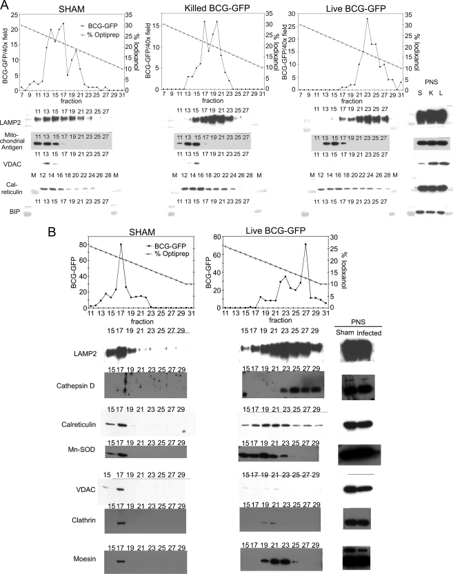
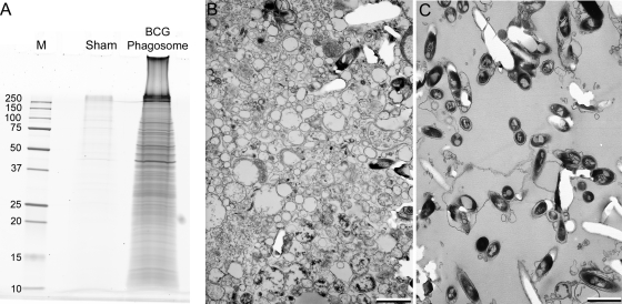
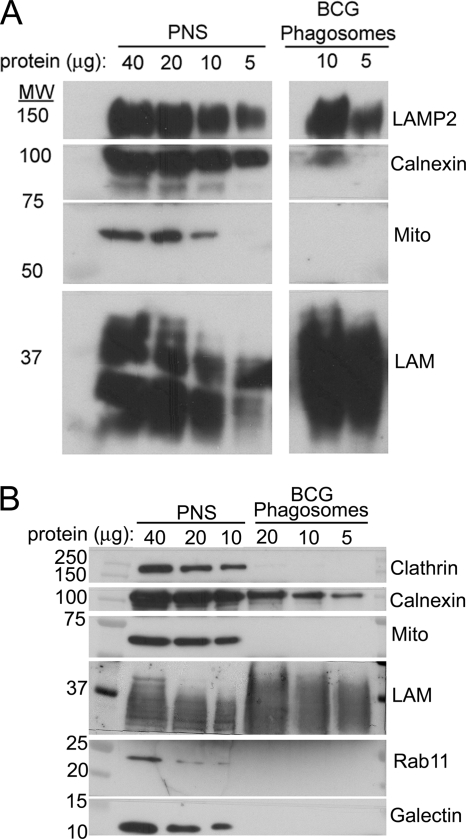
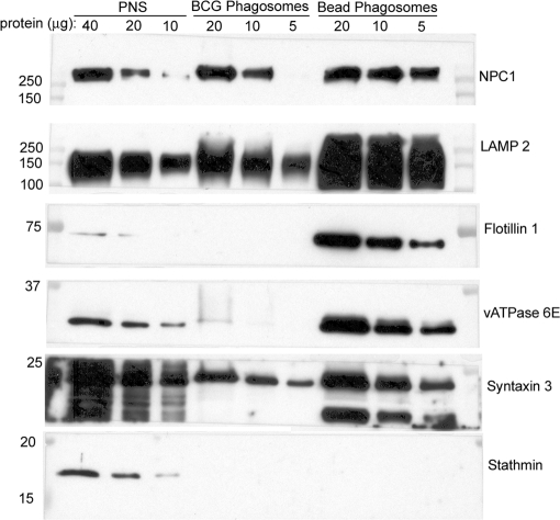
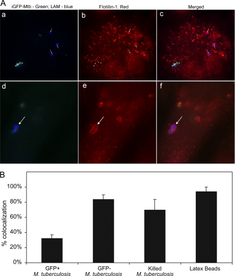
Mycobacterium tuberculosis and Mycobacterium bovis bacille Calmette-Guérin (BCG) alter the maturation of their phagosomes and reside within a compartment that resists acidification and fusion with lysosomes. To define the molecular composition of this compartment, we developed a novel method for obtaining highly purified phagosomes from BCG-infected human macrophages and analyzed the phagosomes by Western immunoblotting and mass spectrometry-based proteomics. Our purification procedure revealed that BCG grown on artificial medium becomes less dense after growth in macrophages. By Western immunoblotting, LAMP-2, Niemann-Pick protein C1, and syntaxin 3 were readily detectable on the BCG phagosome but at levels that were lower than on the latex bead phagosome; flotillin-1 and the vacuolar ATPase were barely detectable on the BCG phagosome but highly enriched on the latex bead phagosome. Immunofluorescence studies confirmed the scarcity of flotillin on BCG phagosomes and demonstrated an inverse correlation between bacterial metabolic activity and flotillin on M. tuberculosis phagosomes. By mass spectrometry, 447 human host proteins were identified on BCG phagosomes, and a partially overlapping set of 289 human proteins on latex bead phagosomes was identified. Interestingly, the majority of the proteins identified consistently on BCG phagosome preparations were also identified on latex bead phagosomes, indicating a high degree of overlap in protein composition of these two compartments. It is likely that many differences in protein composition are quantitative rather than qualitative in nature. Despite the remarkable overlap in protein composition, we consistently identified a number of proteins on the BCG phagosomes that were not identified in any of our latex bead phagosome preparations, including proteins involved in membrane trafficking and signal transduction, such as Ras GTPase-activating-like protein IQGAP1, and proteins of unknown function, such as FAM3C. Our phagosome purification procedure and initial proteomics analyses set the stage for a quantitative comparative analysis of mycobacterial and latex bead phagosome proteomes.


Figures

Fig. 1.
Fig. 1.
Sedimentation of M. bovis BCG phagosomes from THP-1 cells on iodixanol density gradients 3 days postinfection. A, human THP-1 macrophages were allowed to phagocytose live or killed M. bovis BCG-GFP or left untreated (SHAM), and a phagosomal fraction was purified 3 days postinfection. The sham phagosome preparations were processed identically to the other phagosome preparations except that M. bovis BCG was added to the macrophages at the end rather than the start of the incubation period and immediately prior to homogenization. Macrophages from all preparations were homogenized, PNS fractions were obtained by low speed centrifugation, and a phagosomal fraction was obtained by centrifugation through 15% sucrose onto 30% iodixanol. The BCG phagosomes (and free BCG in the case of the sham preparation) were collected from the interface and applied to linear iodixanol gradients. Gradient fractions were collected from the bottom, and aliquots representing equal fractions of the iodixanol gradient or the PNS (0.06% of the entire gradient or 0.06% of the total PNS, respectively) were analyzed by one-dimensional SDS-PAGE and Western immunoblotting. The relative number of BCG-GFP per gradient fraction was determined by mixing equal aliquots of each fraction with formaldehyde and counting the number of green fluorescent bacteria per 400× field by fluorescence microscopy. The BCG phagosomes are lighter than the majority of the mitochondria and ER and go to higher numbered fractions. Whereas LAMP-2 co-sediments with the BCG phagosomes, the mitochondrial antigen markers and the ER markers (calreticulin and BiP) are predominantly found in denser fractions. Whereas the mitochondrial marker VDAC and the ER marker BiP are abundant in the PNS, the majority of these organelles were removed in the purification step prior to the linear iodixanol gradient (low speed sedimentation through 15% sucrose) and thus are present at low levels in the iodixanol gradient fractions. S, sham; K, killed BCG; L, live BCG; M, molecular mass marker lane. B shows an independent replication of the purification of live and sham BCG phagosomes on an iodixanol gradient with analysis of gradient fractions by SDS-PAGE and Western immunoblotting for organelle markers. The BCG phagosomes co-elute with strong bands of LAMP-2 and cathepsin D. As is the case in A, the majority of ER and mitochondria are removed at the step prior to the linear iodixanol gradient. Some residual ER is apparent by calreticulin staining, mostly in fractions that are denser (lower numbered fractions) than the BCG phagosomes, although some calreticulin does co-elute. The mitochondria (stained by the mitochondrial antigens manganese superoxide dismutase (Mn-SOD) and VDAC) also elute in fractions that are denser than the BCG phagosomes. Clathrin (present on plasma membrane) and moesin (a cytoskeletal marker) are abundant in the PNS but are not detected by immunostaining in the peak fractions containing the BCG phagosomes.
Fig. 2.
Fig. 2.
Analysis of BCG phagosomes purified 3 days postinfection from THP-1 cells by SDS-PAGE and electron microscopy. A, BCG phagosomes purified from THP-1 cells 3 days after infection (BCG Phagosome) and sham phagosomes (Sham) prepared from identically treated, non-infected THP-1 cells to which BCG bacteria were added at the end rather than the beginning of the incubation period and immediately prior to homogenization were applied to a one-dimensional SDS-PAGE gradient gel and stained for protein by SYPRO Ruby. Masses of standard molecular mass markers (lane labeled “M”) are indicated in kDa. B and C, transmission electron microscopy of the PNS starting material (pelleted by centrifugation at 10,000 × g for 10 min) (B) and the final purified BCG-GFP phagosomal pellet (C) demonstrates that the purified phagosomes (C) have relatively little contamination and are considerably enriched in BCG phagosomes compared with the PNS starting material (B). Size bars, 1 μm.
Fig. 3.
Fig. 3.
Analysis of 3-day purified BCG phagosomes from THP-1 cells by Western immunoblotting. A, comparison of levels of LAMP-2, calnexin, 60-kDa mitochondrial antigen, and mycobacterial LAM. Samples containing 5–40 μg of protein from the PNS or BCG phagosomes prepared from THP-1 cells 3 days postinfection were analyzed by Western immunoblotting. Whereas LAMP-2 and mycobacterial LAM are enriched on the BCG phagosomes relative to the amount present in the PNS per μg of protein, calnexin is present at a markedly reduced level, and the 60-kDa mitochondrial antigen (Mito) is not detected in the purified BCG phagosome fraction. B, comparison of levels of clathrin, calnexin, mitochondrial antigen (Mito), LAM, Rab-11, and galectin-1. A separate Western immunoblot experiment demonstrates the enrichment of LAM in the BCG phagosome preparation and the persistence of low levels of calnexin relative to the levels present in the PNS. In contrast, clathrin, the mitochondrial antigen, Rab-11, and galectin-1 are detected in the PNS but not in the BCG phagosome preparation.
Fig. 4.
Fig. 4.
Analysis of 3-day purified BCG phagosomes, latex bead phagosomes, and PNS by Western immunoblotting. Samples containing 5–40 μg of protein from the PNS, purified BCG phagosomes, or purified latex bead phagosomes prepared from THP-1 cells 3 days postinfection were analyzed by Western immunoblotting. NPC1, LAMP-2, and syntaxin 3 are enriched on the latex bead phagosomes relative to the levels observed on BCG phagosomes and the PNS. Flotillin-1 and the vATPase are greatly enriched on the latex bead phagosome and relatively scarce on the BCG phagosome. The cytoskeletal protein stathmin, on the other hand, is detected in the PNS but not in the latex bead or BCG phagosome preparations.
Fig. 5.
Fig. 5.
Flotillin-1 immunofluorescence on M. tuberculosis phagosomes in THP-1 cells correlates inversely with metabolic activity of mycobacteria. A, metabolic activity of Mtb-iGFP was assessed by addition of IPTG after infection and visualization of green fluorescent protein expression at fixation 48 h postinfection (a and d), and Mtb-iGFP, independent of metabolic status, was visualized by blue fluorescence with aminomethylcoumarin-labeled anti-LAM antibody (a and d). Flotillin-1 distribution was visualized by staining with Texas Red-labeled anti-human flotillin-1 antibody (b and e). The merged color images are shown on the right (c and f). Metabolically inactive Mtb-iGFP (bacteria that do not express GFP after IPTG induction; a–f, arrows) consistently colocalized with flotillin-1 labeled with Texas Red (arrows indicating three bacteria in b and one bacterium in e). The metabolically active Mtb-iGFP (a–c, arrowheads) showed weaker and less consistent colocalization with flotillin-1 (b). B, quantitative assessment of flotillin-1 immunofluorescence demonstrates relatively low levels of colocalization of flotillin-1 on metabolically active Mtb-iGFP (GFP+ M. tuberculosis) compared with much higher levels of colocalization with inactive Mtb-iGFP (GFP− M. tuberculosis), formalin-killed Mtb-GFP (Killed M. tuberculosis), and latex beads at 48 h postinfection (and 46.5 h post-IPTG induction) in THP-1 cells. The experiment was performed twice with similar results. Values shown are means and error bars indicate S.D. for duplicate determinations for at least 40 bacteria or beads.

Similar articles

Cited by

References

    1. Xu S., Cooper A., Sturgill-Koszycki S., van Heyningen T., Chatterjee D., Orme I., Allen P., Russell D. G. (1994) Intracellular trafficking in Mycobacterium tuberculosis and Mycobacterium avium-infected macrophages. J. Immunol. 153, 2568–2578 - PubMed
    1. Malik Z. A., Denning G. M., Kusner D. J. (2000) Inhibition of Ca(2+) signaling by Mycobacterium tuberculosis is associated with reduced phagosome-lysosome fusion and increased survival within human macrophages. J. Exp. Med. 191, 287–302 - PMC - PubMed
    1. Goren M. B., D’Arcy Hart P., Young M. R., Armstrong J. A. (1976) Prevention of phagosome-lysosome fusion in cultured macrophages by sulfatides of Mycobacterium tuberculosis. Proc. Natl. Acad. Sci. U.S.A. 73, 2510–2514 - PMC - PubMed
    1. Crowle A. J., Dahl R., Ross E., May M. H. (1991) Evidence that vesicles containing living, virulent Mycobacterium tuberculosis or Mycobacterium avium in cultured human macrophages are not acidic. Infect. Immun. 59, 1823–1831 - PMC - PubMed
    1. Armstrong J. A., Hart P. D. (1971) Response of cultured macrophages to Mycobacterium tuberculosis with observations on fusion of lysosomes with phagosomes. J. Exp. Med. 134, 713–740 - PMC - PubMed

Publication types

MeSH terms