. 2010 Feb 1;85(3):444-53.
doi: 10.1093/cvr/cvp335. Epub 2009 Oct 8.

Long-term improvement in mdx cardiomyopathy after therapy with peptide-conjugated morpholino oligomers

Affiliations

Long-term improvement in mdx cardiomyopathy after therapy with peptide-conjugated morpholino oligomers

Natee Jearawiriyapaisarn et al. Cardiovasc Res. .

Abstract

Aims: The cardiomyopathy found in Duchenne muscular dystrophy (DMD) is responsible for death due to heart failure in approximately 30% of patients and additionally contributes to many DMD morbidities. Strategies to bypass DMD-causing mutations to allow an increase in body-wide dystrophin have proved promising, but increasing cardiac dystrophin continues to be challenging. The purpose of this study was to determine if therapeutic restoration of cardiac dystrophin improved the significant cardiac hypertrophy and diastolic dysfunction identified in X-linked muscular dystrophy (mdx) dystrophin-null mouse due to a truncation mutation over time after treatment.

Methods and results: Mice lacking dystrophin due to a truncation mutation (mdx) were given an arginine-rich, cell-penetrating, peptide-conjugated phosphorodiamidate morpholino oligomer (PPMO) that delivered a splice-switching oligonucleotide-mediated exon skipping therapy to restore dystrophin in mdx mice before the development of detectable cardiomyopathy. PPMO successfully restored cardiac dystrophin expression, preserved cardiac sarcolemma integrity, and prevented the development of cardiac pathology that develops in mdx-null mice over time. By echocardiography and Doppler analysis of the mitral valve, we identified that PPMO treatment of mdx mice prevented the cardiac hypertrophy and diastolic dysfunction identified in sham-treated, age-matched mdx mice, characteristic of DMD patients early in the disease process, in as little as 5-6 weeks after the initiation of treatment. Surprisingly, despite the short-term replacement of cardiac dystrophin (<1% present after 12 weeks by immunodetection), PPMO therapy also provided a durable cardiac improvement in cardiac hypertrophy and diastolic dysfunction for up to 7 months after the initiation of treatment.

Conclusion: These results demonstrate for the first time that PPMO-mediated exon skipping therapy early in the course of DMD may effectively prevent or slow down associated cardiac hypertrophy and diastolic dysfunction with significant long-term impact.


Figures

Figure 1
Figure 1
Treatment regimen of PPMOs in mdx mice. Age-matched mdx mice were treated with AVI-5225 or the sham PPMO 623-25-B for two cycles of four once daily i.v. injections at 12 mg/kg/day. Two groups were studied to determine short-term effects of AVI-5225 therapy on the development or mdx cardiomyopathy (Group 1, top) and the long-term effects of AVI-5225 therapy (Group 2, bottom). The effects of short-term treatment were determined at 5 and 6 weeks after the initiation of treatment (Group 1, top). Long-term effects were determined after ∼7 months after the initiation of treatment (Group 2, bottom). Serum was collected at indicated time points to monitor the level of creatine kinase (CK) and CK-MB isoenzyme. RNA, protein. Histology of cardiac muscle was analysed 6 weeks after the initiation of therapy. Cardiac metrics mice were analysed by echocardiography (Echo) and Doppler analysis at baseline, 5 weeks (Group 1), or ∼28 weeks (Group 2) after the initiation of therapy. *Echo and Doppler measurements were performed on independent age-matched mice and compared with those measured at later timepoints.
Figure 2
Figure 2
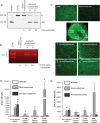
Global dystrophin expression in the heart of mdx mice induced by PPMO AVI-5225 prevents the loss of sarcolemma integrity 6 weeks after the initiation of therapy. (A) The level of exon 23-skipped mRNA was analysed by nested RT–PCR 3 weeks after the last injection. The upper band (445 bp) indicated by FL corresponds to the full-length dystrophin transcript, and the lower band (232 bp) indicated by Δ23 corresponds to the exon 23-skipped dystrophin transcript. The percentages of exon 23 skipping are shown. Samples A and B are from different mice. (B) In-gel western detection of total protein extracted from injected mdx heart. Dystrophin (DYS) was detected by the NCL-DYS2 monoclonal antibody. The percentages of dystrophin restoration compared with the average of two different wild-type mice are shown. (C) Immunofluorescence detection of dystrophin in the treated mdx heart 6 weeks after the start of therapy. Panels 1–4 are higher-magnification images of the regions shown in the full-view image of the AVI-5225- treated heart. Panel 1: septum; 2: anterior left ventricle wall (LV); 3: posterior LV; 4: lateral LV. C57BL: heart from normal C57BL mouse; Sham-treated: heart from PPMO 623-25-B-treated mdx mouse. Scale bar, 100 µm. Significantly less serum CK (D) and CK-MB (E) is circulating in mdx mice after AVI-5225 treatment compared with age-matched mdx sham-treated controls. C57BL (n = 10), Sham-treated mdx mice (n = 11), and AVI-5225-treated mdx mice (n = 6). A one-way ANOVA was performed to determine significance, followed by a Holm-Sidak pairwise comparison to significance between groups, *P < 0.05 for comparisons to wild-type C57BL; †P < 0.05 for comparisons to sham-treated mdx mice.
Figure 3
Figure 3
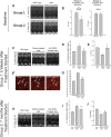
Treatment of mdx mice with AVI-5225 prevents the development of mdx-associated cardiac hypertrophy and diastolic dysfunction, which leads to significant improvements in long-term durability. No differences in cardiac wall thickness shown by representative M mode (A) or diastolic function determined by mitral valve Doppler analysis (E/A ratio) (B) are present in mdx and wild-type control mice at ages 8 or 16 weeks old. n.s. = not significant. (C) AVI-5225 treatment prevents the development of anterior and posterior wall thickening in mdx mice as evidence by representative M-mode; (D) LV Mass determined by echocardiography, *P < 0.001 vs. wild type; †P < 0.001 vs. sham-treated mdx mice; and (E) prevents the development of diastolic dysfunction in mdx hearts determined by mitral valve Doppler analysis, *P < 0.05 vs. wild-type; †P < 0.05 vs. sham-treated mdx. (F) Representative cross sectional micrographs of wild-type (left, n = 3), sham-treated mdx (middle, n = 3), and AVI-5225-treated mdx mice (right, n = 3) hearts, illustrating that cardiomyocytes from AVI-5225-treated mdx mice do not develop cardiomyocyte hypertrophy 5 weeks after the initiation of therapy. Arrows indicate representative cross-sectional areas. Scale bar, 10 µm. (G) Quantitative analysis of cardiomyocyte cross-sectional areas demonstrates this protection. n = 3 mice/group; each mean is calculated from 50 measurements from multiple sections from the three representative mice from each group. *P < 0.001 vs. wild-type; †P < 0.001 vs. sham-treated mdx mice. (H) Durable improvement in mdx mice ∼7 months after AVI-5225 was identified by reduced anterior and posterior wall thickening as evidenced by representative M-mode; (I) LV mass determined by echocardiography, *P < 0.05 vs. wild-type; †P < 0.01 vs. sham-treated mdx. (J) Residual improvement in diastolic dysfunction determined by mitral valve Doppler analysis was identified as not statistically significant between wild-type or sham-treated mdx groups. *P < 0.05 vs. wild-type. A one-way ANOVA was performed to determine significance, followed by a Holm-Sidak pairwise comparison to significance between groups, with comparison groups identified above.
Figure 4
Figure 4
Treatment of mdx mice 6 weeks after the initiation of therapy does not alter the mild rare focal fibrosis. Representative heart sections from wild-type (A and B), sham-treated mdx (C and D), and AVI-5225-treated mdx (E and F) mice were analysed after Trichrome (A, C, E) or H&E (B, D, F) staining (20×). While rare focal fibrosis is present in both mdx and AVI-5225-treated mdx mice, all three groups of mice are generally indistinguishable by histology.

Comment in

References

    1. Monaco AP, Bertelson CJ, Liechti-Gallati S, Moser H, Kunkel LM. An explanation for the phenotypic differences between patients bearing partial deletions of the DMD locus. Genomics. 1988;2:90–95. - PubMed
    1. Bauman J, Jearawiriyapaisarn N, Kole R. Therapeutic potential of splice-switching oligonucleotides. Oligonucleotides. 2009;19:1–14. - PMC - PubMed
    1. van Deutekom JC, Janson AA, Ginjaar IB, Frankhuizen WS, Aartsma-Rus A, Bremmer-Bout M, et al. Local dystrophin restoration with antisense oligonucleotide PRO051. N Engl J Med. 2007;357:2677–2686. - PubMed
    1. Dunckley MG, Manoharan M, Villiet P, Eperon IC, Dickson G. Modification of splicing in the dystrophin gene in cultured Mdx muscle cells by antisense oligoribonucleotides. Hum Mol Genet. 1998;7:1083–1090. - PubMed
    1. Wilton SD, Lloyd F, Carville K, Fletcher S, Honeyman K, Agrawal S, et al. Specific removal of the nonsense mutation from the mdx dystrophin mRNA using antisense oligonucleotides. Neuromuscul Disord. 1999;9:330–338. - PubMed

Publication types