Comparative Study
. 2009 Dec;297(6):G1085-92.
doi: 10.1152/ajpgi.00119.2009. Epub 2009 Oct 8.

Cholecystokinin-58 and cholecystokinin-8 exhibit similar actions on calcium signaling, zymogen secretion, and cell fate in murine pancreatic acinar cells

Affiliations
Comparative Study

Cholecystokinin-58 and cholecystokinin-8 exhibit similar actions on calcium signaling, zymogen secretion, and cell fate in murine pancreatic acinar cells

David N Criddle et al. Am J Physiol Gastrointest Liver Physiol. 2009 Dec.

Abstract

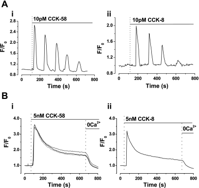
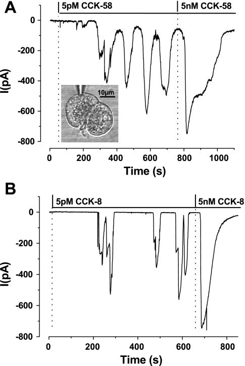
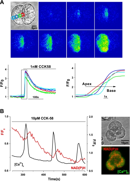
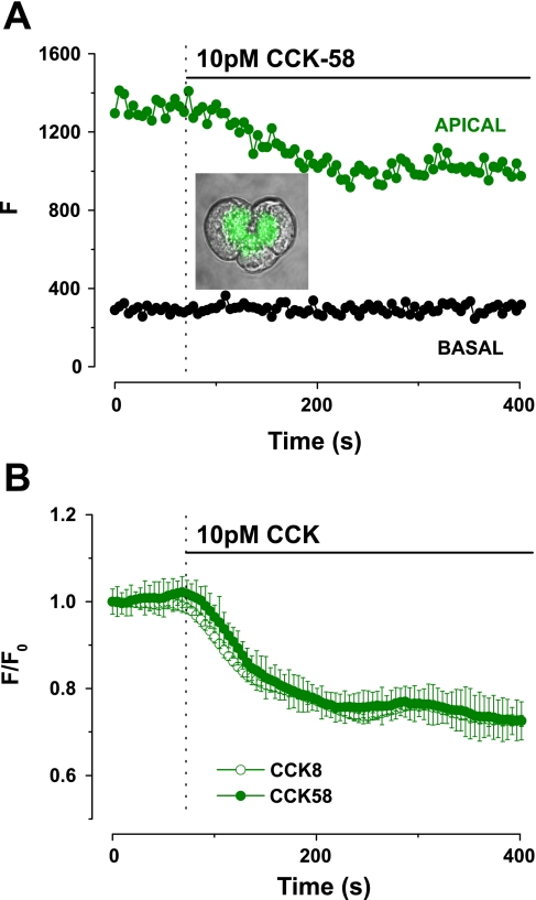
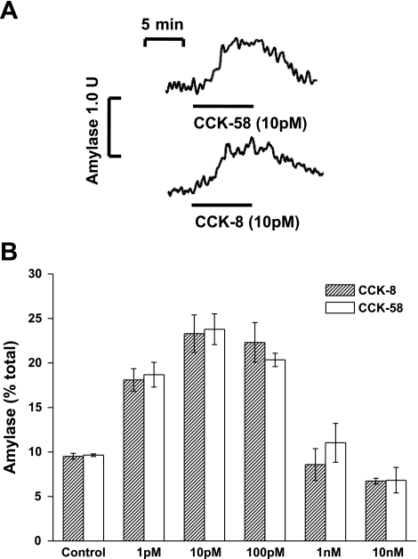
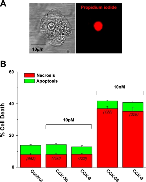
The gastrointestinal hormone CCK exists in various molecular forms, with differences in bioactivity between the well-characterized CCK-8 and larger CCK-58 previously reported. We have compared the effects of these peptides on cytosolic calcium concentration ([Ca(2+)](c)), mitochondrial metabolism, enzyme secretion, and cell fate in murine isolated pancreatic acinar cells using fluorescence confocal microscopy and patch-clamp electrophysiology. CCK-58 (1-10 pM) induced transient, oscillatory increases of [Ca(2+)](c), which showed apical to basolateral progression and were associated with a rise of mitochondrial NAD(P)H. CCK-58 (10 pM) induced zymogen exocytosis in isolated cells and amylase secretion from isolated cells and whole tissues. Hyperstimulation with supraphysiological CCK-58 (5 nM) induced a single large increase of [Ca(2+)](c) that declined to a plateau, which remained above the basal level 20 min after application and was dependent on external Ca(2+) entry. In cells dispersed from the same tissues, CCK-8 induced similar patterns of responses to those of CCK-58, with oscillatory increases of [Ca(2+)](c) at lower (pM) concentrations and sustained responses at 5 nM. CCK-58 and CCK-8 exhibited similar profiles of action on cell death, with increases in necrosis at high CCK-58 and CCK-8 (10 nM) that were not significantly different between peptides. The present experiments indicate that CCK-8 and CCK-58 have essentially identical actions on the acinar cell at high and low agonist concentrations, suggesting an action via the same receptor and that the differences observed in an intact rat model may result from indirect effects of the peptides. Our data strengthen the argument that CCK-58 is an important physiological form of this gastrointestinal hormone.


Figures

Fig. 1.
Fig. 1.
Comparative effects of low (pM) and high (nM) CCK-58 and CCK-8 on cytosolic calcium concentration ([Ca2+]c) in isolated murine pancreatic acinar cells. Representative traces show that application of 10 pM (A) CCK-58 (i) and CCK-8 (ii) induced oscillatory rises of [Ca2+]c, whereas application of 5 nM (B) CCK-58 (i) (recordings from 3 cells shown) and CCK-8 (ii) evoked single “spike” increases in [Ca2+]c, followed by plateau sustained elevations above the basal level. When extracellular Ca2+ (with EGTA) was removed from the perfusion solution, the plateau increase in [Ca2+]c was reversed to basal levels, indicating the dependence on Ca2+ influx into the cell. F0 represents the initial fluorescence recorded at the start of the experiment, and F represents the fluorescence recorded at specific time points.
Fig. 2.
Fig. 2.
Typical recordings obtained from whole cell patch-clamp experiments, contrasting the effects of CCK-58 (A) (5 pM-5 nM, n = 5) with CCK-8 (B) (5 pM-5 nM, n = 6) to induce Ca2+-dependent chloride currents (IClCa) in isolated pancreatic acinar cells. Cells were held at −30 mV, and CCK was applied once the membrane had been ruptured mechanically by suction. At the lower (5 pM) concentration both CCK-58 and CCK-8 induced oscillatory inward IClCa currents, denoting rises of [Ca2+]c, whereas at 5 nM a larger, broader single current was observed. The inset in A shows a light-transmitted image of a doublet of pancreatic acinar cells with the patch-pipette recording from the upper cell.
Fig. 3.
Fig. 3.
Characteristics of CCK-58-induced elevations of [Ca2+]c in isolated pancreatic acinar cells. A: typical fluorescent images and traces showing the apical to basal progression of the rise in [Ca2+]c recorded in 1 of a doublet of acinar cells stimulated by CCK-58 (1 nM, n = 6). Regions of interest for measurement of the apical (red) to basal (blue) [Ca2+]c signals are shown in the inset light-transmitted image. B: typical trace showing the stimulus-metabolism coupling induced by 10 pM CCK-58. After each elevation of [Ca2+]c induced by CCK-58, a slower rise of NAD(P)H (red) was observed. The perigranular distribution of the mitochondrial belt can be seen clearly by the NAD(P)H (red) in the inset fluorescent image of an acinar cell triplet. Similar responses to 10 pM CCK-8 were observed in separate experiments (n = 6).
Fig. 4.
Fig. 4.
Comparative effects of CCK-58 and CCK-8 on digestive enzyme secretion in murine pancreatic acinar cells. A: typical trace showing exocytosis that occurred in response to a physiological concentration (10 pM) of CCK-58. Typical quinacrine staining (green) of zymogen granules is shown within the inset light-transmitted image. On stimulation with CCK-58 apical quinacrine fluorescence (F) fell from a steady baseline to a plateau level, with no change in the basal area. B: exocytotic responses induced by CCK-58 (n = 16) and CCK-8 (n = 11) were not significantly different. All fluorescence changes were normalized from basal values (F/F0).
Fig. 5.
Fig. 5.
A: typical traces showing the reversible amylase secretion in response to CCK-58 and CCK-8 from superfused segments of murine pancreatic tissue, monitored fluorometrically in the flow cell effluent (n = 5). CCK-58 and CCK-8 increased amylase secretion to 0.98 and 0.80 U/min per 100 mg of tissue from basal levels of 0.55 and 0.49 U/min per 100 mg of tissue, respectively. B: concentration-dependent effects of CCK-58 (n = 3) and CCK-8 (n = 3) (1 pM-10 nM) on amylase secretion from isolated murine pancreatic cells, expressed as a percentage of total amylase. No significant difference between CCK-58 and CCK-8 responses was detected at all concentrations tested (P > 0.05).
Fig. 6.
Fig. 6.
Comparative effects of CCK-58 and CCK-8 on cell death. A: light-transmitted and propidium iodide fluorescent images showing bright staining of the nucleus of a necrotic cell after exposure to 10 nM CCK-58 for 30 min at 37°C. B: mean data from such experiments comparing the effects of CCK-58 and CCK-8 on apoptosis (green) and necrosis (red). No significant increase in total cell death was observed with physiological (10 pM) CCK-58 or CCK-8 stimulation. However, total cell death was increased by hyperstimulation with 10 nM of either peptide. Necrosis significantly increased in response to 10 nM CCK-58 or CCK-8, whereas apoptosis remained at control levels. No significant difference between CCK-58 and CCK-8 was detected in these experiments. (Bars show mean percentage of cell death ± SE. The number of cells analyzed in each group are in parentheses.)
Fig. 7.
Fig. 7.
Comparative effects of CCK-58 and CCK-8 on cell membrane damage and intracellular trypsin activation. A: i: light-transmitted images showing blebbing of the plasmalemmal membrane induced by stimulation with 20 nM CCK-58 for 20 min at 37°C, which increased with exposure time. ii: Mean data comparing the effects of CCK-58 (n = 17) and CCK-8 (n = 10) in this experimental protocol show that there was no significant difference detected between the detrimental actions of these peptides. (Bars show mean levels of cell death ± SE. The number of cells analyzed in each group are in parentheses.) B: light-transmitted image and rhodamine 110, bis-(CBZ-L-isoleucyl-L-prolyl-L-arginine amide) dihydrochloride (BZiPAR) fluorescent images of a doublet of acinar cells damaged by exposure to 20 nM CCK-58 for 20 min at 7°C (i). Trypsin activity (blue) can be seen at various time points, following bath application of BZiPAR (10 nM) at t = 0 s, commencing in the granular, apical pole of the cell (t = 2 s), and spreading to the basolateral region. Similar trypsin activity was detected in response to both hyperstimulation with CCK-58 (6 of 6 cells) and CCK-8 (6 of 6 cells) but not by stimulation with 10 pM CCK-58 (ii) for the same period (9 of 9 cells).

References

    1. Bi Y, Page SL, Williams JA. Rho and Rac promote acinar morphological changes, actin reorganization, and amylase secretion. Am J Physiol Gastrointest Liver Physiol 289: G561–G570, 2005 - PubMed
    1. Campos-Toimil M, Edwardson JM, Thomas P. Real-time studies of zymogen granule exocytosis in intact rat pancreatic acinar cells. J Physiol 528: 317–326, 2000 - PMC - PubMed
    1. Criddle DN, Gerasimenko JV, Baumgartner HK, Jaffar M, Voronina S, Sutton R, Petersen OH, Gerasimenko OV. Calcium signalling and pancreatic cell death: apoptosis or necrosis? Cell Death Differ 14: 1285–1294, 2007 - PubMed
    1. Criddle DN, Gillies S, Baumgartner-Wilson HK, Jaffar M, Chinje EC, Passmore S, Chvanov M, Barrow S, Gerasimenko OV, Tepikin AV, Sutton R, Petersen OH. Menadione-induced reactive oxygen species generation via redox cycling promotes apoptosis of murine pancreatic acinar cells. J Biol Chem 281: 40485–40492, 2006 - PubMed
    1. Criddle DN, Murphy J, Fistetto G, Barrow S, Tepikin AV, Neoptolemos JP, Sutton R, Petersen OH. Fatty acid ethyl esters cause pancreatic calcium toxicity via inositol trisphosphate receptors and loss of ATP synthesis. Gastroenterology 130: 781–793, 2006 - PubMed

Publication types

MeSH terms

LinkOut - more resources