. 2009 Dec;297(6):G1093-106.
doi: 10.1152/ajpgi.00292.2009. Epub 2009 Oct 8.

Hedgehog pathway activation and epithelial-to-mesenchymal transitions during myofibroblastic transformation of rat hepatic cells in culture and cirrhosis

Affiliations

Hedgehog pathway activation and epithelial-to-mesenchymal transitions during myofibroblastic transformation of rat hepatic cells in culture and cirrhosis

Steve S Choi et al. Am J Physiol Gastrointest Liver Physiol. 2009 Dec.

Abstract

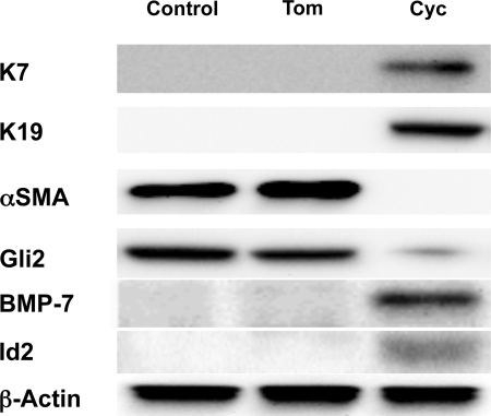
Myofibroblastic hepatic stellate cells (MF-HSC) are derived from quiescent hepatic stellate cells (Q-HSC). Q-HSC express certain epithelial cell markers and have been reported to form junctional complexes similar to epithelial cells. We have shown that Hedgehog (Hh) signaling plays a key role in HSC growth. Because Hh ligands regulate epithelial-to-mesenchymal transition (EMT), we determined whether Q-HSC express EMT markers and then assessed whether these markers change as Q-HSC transition into MF-HSC and whether the process is modulated by Hh signaling. Q-HSC were isolated from healthy livers and cultured to promote myofibroblastic transition. Changes in mRNA and protein expression of epithelial and mesenchymal markers, Hh ligands, and target genes were monitored in HSC treated with and without cyclopamine (an Hh inhibitor). Studies were repeated in primary human HSC and clonally derived HSC from a cirrhotic rat. Q-HSC activation in vitro (culture) and in vivo (CCl(4)-induced cirrhosis) resulted in decreased expression of Hh-interacting protein (Hhip, an Hh antagonist), the EMT inhibitors bone morphogenic protein (BMP-7) and inhibitor of differentiation (Id2), the adherens junction component E-cadherin, and epithelial keratins 7 and 19 and increased expression of Gli2 (an Hh target gene) and mesenchymal markers, including the mesenchyme-associated transcription factors Lhx2 and Msx2, the myofibroblast marker alpha-smooth muscle actin, and matrix molecules such as collagen. Cyclopamine reverted myofibroblastic transition, reducing mesenchymal gene expression while increasing epithelial markers in rodent and human HSC. We conclude that Hh signaling plays a key role in transition of Q-HSC into MF-HSC. Our findings suggest that Q-HSC are capable of transitioning between epithelial and mesenchymal fates.


Figures

Fig. 1.
Fig. 1.
Changes in expression of epithelial and mesenchymal genes during culture-induced transition of primary rat quiescent hepatic stellate cells (Q-HSC) into myofibroblastic hepatic stellate cells (MF-HSC). Primary HSC were isolated from 6 healthy adult male rats per experiment, pooled, and cultured on plastic dishes in serum-containing medium. RNA was isolated at different time points, and changes in gene expression were evaluated by quantitative RT-PCR (qRT-PCR). A: myofibroblastic markers [α-smooth muscle actin (α-SMA), collagen 1α1 (Col1α1), and fibronectin]. B: markers of quiescent HSC [peroxisome proliferator-activated receptor-γ (PPARγ) and glial fibrillary acidic protein (GFAP)]. C: epithelial markers [keratin 7 (K7), keratin 19 (K19), and desmoplakin]. Values are means ± SE of triplicate experiments. *P < 0.05; **P < 0.01; †P < 0.005.
Fig. 2.
Fig. 2.
Comparison of epithelial and mesenchymal gene expression in normal rat cholangiocytes (NRC) and primary rat stellate cells. Primary HSC were isolated from healthy adult male rats and cultured on plastic dishes in serum-containing medium. NRC were cultured on collagen-coated dishes in serum-supplemented medium. RNA was isolated from NRC and quiescent and culture-activated HSC (day 7), with changes in gene expression evaluated by qRT-PCR. A: HSC-associated markers (Lhx2, Msx2, and desmin). B: markers of quiescent HSC (PPARγ and GFAP). C: epithelial markers (K7 and K19). D: factors that inhibit EMT [bone morphogenetic protein-7 (BMP-7), inhibitor of differentiation (Id2), and desmoplakin]. Values are means ± SE of triplicate experiments. *P < 0.05; **P < 0.01; †P < 0.005.
Fig. 3.
Fig. 3.
Culture-related changes in HSC expression of genes that regulate epithelial-to-mesenchymal transitions (EMT) determined by qRT-PCR analysis of RNA obtained from the primary rat HSC described in Fig. 1. A: factors that inhibit EMT (BMP-7, Id2, and E-cadherin). B: factors that promote EMT [Snail, transforming growth factor-β (TGF-β), and S100A4]. C: Hedgehog (Hh) signaling factors known to regulate EMT [Hedgehog-interacting protein (Hhip), Sonic Hh (Shh), and Gli2]. Values are means ± SE of triplicate experiments. *P < 0.05; **P < 0.01; †P < 0.005.
Fig. 4.
Fig. 4.
Culture-induced changes in protein expression of EMT-related factors during transition of Q-HSC into MF-HSC. A: Oil Red O staining to detect neutral lipids and immunocytochemistry for K19, an epithelial keratin, in a representative, freshly isolated HSC from preparations described in Fig. 1. Immunocytochemistry in NRC confirms K19 positivity. Magnification ×40. B: Western blot analysis of protein harvested from these primary rat HSC. Results are representative of triplicate experiments.
Fig. 5.
Fig. 5.
Effects of inhibiting Hh signaling on mRNA expression of epithelial and mesenchymal markers in cultured primary rat HSC. Primary stellate cells were isolated from another 2 healthy adult male rats per experiment, pooled, and cultured on plastic in serum-containing medium for 4 days. Then cyclopamine (Cyc, a pharmacological inhibitor of Hh signaling) or tomatidine (Tom, an inert cyclopamine analog) was added, and cultures were harvested after an additional 4 or 7 days. RNA was isolated, and changes in gene expression were monitored by qRT-PCR. A: myofibroblastic markers (α-SMA, Col1α1, and S100A4). B: epithelial markers (K7, K19, and desmoplakin). C: EMT-inhibitory markers (E-cadherin and BMP-7). Values are means ± SE of triplicate experiments. *P < 0.05.
Fig. 6.
Fig. 6.
Effects of inhibiting Hh signaling on protein expression of representative epithelial and mesenchymal markers in cultured primary rat HSC. Whole cell protein was isolated from HSC cultures described in Fig. 5. Changes in protein expression of K7, K19, α-SMA, and BMP-7 were assessed by Western blot analysis (40 μg whole cell protein/lane). β-Actin was used as a loading control.
Fig. 7.
Fig. 7.
Changes in mRNA expression of mesenchymal markers in cultured human HSC during myofibroblastic transition and effects of Hh pathway modulation. A and B: primary HSC were harvested from a residual ssegment of healthy human liver tissue that was used for split-liver transplantation. HSC were culture activated to MF-HSC on plastic dishes in serum-containing medium. qRT-PCR was done to compare gene expression in Q-HSC and MF-HSC (A) and after MF-HSC were treated with tomatidine or cyclopamine for 4 days (B). C: effects of similar treatment with tomatidine or cyclopamine on gene expression in the clonal human MF-HSC line LX-2. Values are means ± SE of triplicate experiments. *P < 0.05; †P < 0.005.
Fig. 8.
Fig. 8.
Effects of Hh pathway inhibition on expression of EMT-related genes in primary human MF-HSC. Primary human HSC described in Fig. 7 were treated with tomatidine or cyclopamine, and gene expression changes were compared by qRT-PCR. A: Hh signaling pathway factors (Gli1 and Gli2) and EMT inhibitors (BMP-7 and Id2). B: EMT promoters (Snail, Slug, and S100A4). C: epithelial markers (E-cadherin and desmoplakin) and the HSC quiescence marker PPARγ. D: myofibroblastic markers [α-SMA, Col1α1, and plasminogen activator inhibitor-1 (PAI-1)]. Values are means ± SE of triplicate experiments. *P < 0.05; **P < 0.01.
Fig. 9.
Fig. 9.
Effects of Hh signaling inhibition on protein expression of representative epithelial and mesenchymal markers in cultured primary human HSC. Whole cell protein was isolated from HSC cultures described in Fig. 7. Changes in protein expression of K7, K19, α-SMA, Gli2, BMP-7, and Id2 were assessed by Western blot (40 μg whole cell protein/lane). β-Actin was used as a loading control.
Fig. 10.
Fig. 10.
Changes in expression of genes that regulate epithelial-to-mesenchymal transitions (EMT) and Hh pathway activation during CCl4-induced cirrhosis. Liver RNA was isolated from mice that had been treated with CCl4 for 8 wk to induce cirrhosis and compared with age- and sex-matched littermates treated with corn oil vehicle (n = 6 each). qRT-PCR was used to assess expression of Hh signaling factors known to regulate EMT. A: Shh, Gli2, and Hh-interacting protein (Hhip). B: epithelial genes that inhibit EMT (desmoplakin, BMP-7, and Id2). C: factors that promote EMT (TGF-β and S100A4). *P < 0.05.
Fig. 11.
Fig. 11.
Evidence for Hh pathway activation and EMT in clonally derived MF-HSC from livers with CCl4-induced cirrhosis. Clonally derived HSC lines (8B and 5H) from a single rat with CCl4-induced cirrhosis were compared with freshly isolated primary rat Q-HSC to assess markers associated with EMT and Hh signaling. A: mesenchymal marker expression (α-SMA, Col1α1, fibronectin, vimentin, and S100A4). B: Hh signaling (Shh and Gli2). C: epithelial markers (K7, desmoplakin, and E-cadherin). D: EMT markers (BMP-7 and Id2). For each gene, expression was normalized to that of a housekeeping gene (S9) in the same RNA sample and then expressed relative to expression of the same gene in primary Q-HSC. 8B were then treated with tomatidine or cyclopamine, and effects on gene expression changes were assessed by qRT-PCR. E: EMT-inhibitory markers (BMP-7 and desmoplakin) and the Q-HSC marker PPARγ. F: myofibroblastic markers (α-SMA, Col1α1, and S100A4). Values are means ± SE of triplicate experiments. *P < 0.05; **P < 0.01.

References

    1. Acloque H, Adams MS, Fishwick K, Bronner-Fraser M, Nieto MA. Epithelial-mesenchymal transitions: the importance of changing cell state in development and disease. J Clin Invest 119: 1438–1449, 2009 - PMC - PubMed
    1. Asahina K, Tsai SY, Li P, Ishii M, Maxson RE, Jr, Sucov HM, Tsukamoto H. Mesenchymal origin of hepatic stellate cells, submesothelial cells, and perivascular mesenchymal cells during mouse liver development. Hepatology 49: 998–1011, 2009 - PMC - PubMed
    1. Bailey JM, Singh PK, Hollingsworth MA. Cancer metastasis facilitated by developmental pathways: Sonic hedgehog, Notch, and bone morphogenic proteins. J Cell Biochem 102: 829–839, 2007 - PubMed
    1. Choi SS, Sicklick JK, Ma Q, Yang L, Huang J, Qi Y, Chen W, Li YX, Goldschmidt-Clairmont P, Diehl AM. Sustained activation of Rac-1 in hepatic stellate cells promotes liver injury and fibrosis in mice. Hepatology 44: 1267–1277, 2006 - PubMed
    1. De Minicis S, Seki E, Uchinami H, Kluwe J, Zhang Y, Brenner DA, Schwabe RF. Gene expression profiles during hepatic stellate cell activation in culture and in vivo. Gastroenterology 132: 1937–1946, 2007 - PubMed

Publication types

MeSH terms

LinkOut - more resources