. 2009 Nov;175(5):2159-70.
doi: 10.2353/ajpath.2009.090391. Epub 2009 Oct 8.

Contrasting actions of selective inhibitors of angiopoietin-1 and angiopoietin-2 on the normalization of tumor blood vessels

Affiliations

Contrasting actions of selective inhibitors of angiopoietin-1 and angiopoietin-2 on the normalization of tumor blood vessels

Beverly L Falcón et al. Am J Pathol. 2009 Nov.

Abstract

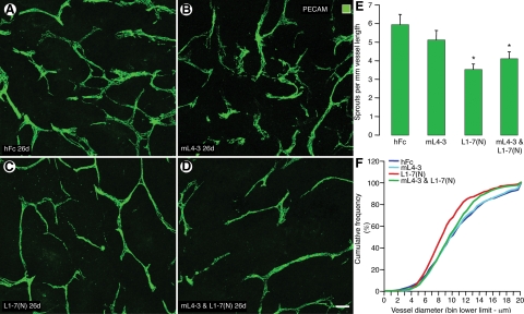
Angiopoietin-1 (Ang1) and angiopoietin-2 (Ang2) have complex actions in angiogenesis and vascular remodeling due to their effects on Tie2 receptor signaling. Ang2 blocks Ang1-mediated activation of Tie2 in endothelial cells under certain conditions but is a Tie2 receptor agonist in others. We examined the effects of selective inhibitors of Ang1 (mL4-3) or Ang2 (L1-7[N]), alone or in combination, on the vasculature of human Colo205 tumors in mice. The Ang2 inhibitor decreased the overall abundance of tumor blood vessels by reducing tumor growth and keeping vascular density constant. After inhibition of Ang2, tumor vessels had many features of normal blood vessels (normalization), as evidenced by junctional accumulation of vascular endothelial-cadherin, junctional adhesion molecule-A, and platelet/endothelial cell adhesion molecule-1 in endothelial cells, increased pericyte coverage, reduced endothelial sprouting, and remodeling into smaller, more uniform vessels. The Ang1 inhibitor by itself had little noticeable effect on the tumor vasculature. However, when administered with the Ang2 inhibitor, the Ang1 inhibitor prevented tumor vessel normalization, but not the reduction in tumor vascularity produced by the Ang2 inhibitor. These findings are consistent with a model whereby inhibition of Ang2 leads to normalization of tumor blood vessels by permitting the unopposed action of Ang1, but decreases tumor vascularity primarily by blocking Ang2 actions.


Figures

Figure 1
Figure 1
Differences in tumor growth and vascularity after inhibition of Ang1 and/or Ang2. A: Growth of Colo205 tumors treated for 26 days with one of four treatment regimens. Growth rates were similar with hFc and mL4-3 (Ang1 inhibitor) but were significantly slower with L1-7(N) (Ang2 inhibitor) or the combination of inhibitors. *P < 0.01 vs. hFc, **P < 0.001 vs. hFc. B: Colo205 tumors stained with the nuclear dye, YO-PRO-1, to show the size of the tumors (green). The tumor treated with the Ang2 inhibitor or the combination of inhibitors was smaller than after the other treatments. C–F. Confocal images showing PECAM immunoreactivity (red) of blood vessels surrounded by YO-PRO-1 staining (green) of viable tumor cells in Colo205 tumors treated for 26 days. The vascular density in YO-PRO-1-positive regions of tumors was similar in control tumors (C, hFc), after inhibition of Ang1 (D, mL4-3), or inhibition of Ang2 (E, L1-7[N]), but was less after inhibition of Ang1 and Ang2 together (F). The area density of PECAM-positive blood vessels in YO-PRO-1–positive regions was not changed by either inhibitor alone but was significantly less after the combination of inhibitors (G). Overall tumor vascular mass, calculated as the product of the fractional area of PECAM staining and tumor size, was significantly less than control after inhibition of Ang2 or after inhibition of Ang1 and Ang2 together (H). *P < 0.05 compared with hFc. Scale bar: 3.5 mm (B); 80 μm (CF).
Figure 2
Figure 2
Tumor vessel phenotype after inhibition of Ang1 and/or Ang2. Confocal microscopic images of endothelial cells (PECAM; green) in Colo205 tumors after treatment for 26 days. Blood vessels are tortuous and sprouting in a control tumor (A, hFc) and after inhibition of Ang1 (B, mL4-3), but are more uniform in size and have less sprouting after inhibition of Ang2 (C, L1-7[N]). Tumor vessels are less numerous after treatment with both inhibitors (D). Endothelial sprouts were significantly less numerous after inhibition of Ang2 (E). This reduction in sprouts by the Ang2 inhibitor was not blocked by co-administration of the Ang1 inhibitor (E). Graph of the size distributions of tumor vessels in the four groups shows that the average size of tumor vessels was significantly less after inhibition of Ang2. Inhibition of Ang1 reduced this effect (F). *P < 0.05 compared with hFc. Scale bar: 50 μm.
Figure 3
Figure 3
Distribution of VE-cadherin and JAM-A after inhibition of Ang1 and/or Ang2. Fluorescence microscopic images of Colo205 tumors stained for the endothelial adherens junction protein VE-cadherin or the tight junction protein JAM-A after treatment for 26 days. VE-cadherin immunoreactivity was weak in control tumors (A, hFc) and after inhibition of Ang1 (B, mL4-3) but was strong and linear at endothelial cell borders after inhibition of Ang2 (C, L1-7(N)). The linear pattern was not present after inhibition of Ang1 and Ang2 (D). Immunoreactivity for the endothelial tight junction protein JAM-A was faint after hFc (E), Ang1 inhibition (F), or inhibition of Ang1 and Ang2 (H), but was strong after inhibition of Ang2 (G). Scale bar = 8 μm.
Figure 4
Figure 4
Distribution of PECAM after inhibition of Ang1 and/or Ang2. Confocal microscopic images showing the distribution of PECAM immunoreactivity in blood vessels of Colo205 tumors treated for 26 days. PECAM staining was strong in all groups (A−D), but was patchy in control tumors (A, hFc), after inhibition of Ang1 (B, mL4-3), or after inhibition of Ang1 and Ang2 (D). By comparison, PECAM staining was largely localized to endothelial cell junctions after inhibition of Ang2 (C, L1-7(N)). Contiguous linear regions of PECAM staining, identified by the algorithm used to measure junctional normalization, are marked by colored lines (E–H). Linear PECAM staining were less in control tumors (E) and after inhibition of Ang1 (F) or inhibition of Ang1 and Ang2 (H) than after inhibition of Ang2 alone (G). Measurements of contiguous linear PECAM staining revealed significantly larger values after inhibition of Ang2 alone than after inhibition of Ang1 and Ang2 (I). *P < 0.05 compared with hFc. **P < 0.05 compared with Ang2 inhibitor. Scale bar = 10 μm.
Figure 5
Figure 5
Pericyte distribution after inhibition of Ang1 and/or Ang2. Fluorescence microscopic images of endothelial cells (PECAM; green) and pericytes (PDGFR-β; red) of blood vessels in Colo205 tumors treated for 26 days. Pericytes were loosely associated with blood vessels in control tumors (A, hFc), after inhibition of Ang1 (B, mL4-3), or after inhibition of Ang1 and Ang2 (D) but were more closely associated with endothelial cells after inhibition of Ang2 (C, L1-7(N)). Pericytes in contact with tumor vessels were sparse and had faint PDGFR-β immunoreactivity after hFc (E), after inhibition of Ang1 (F), or after inhibition of Ang1 and Ang2 (H) but were abundant and had strong PDGFR-β immunoreactivity after inhibition of Ang2 (G). Measurements confirmed the greater abundance of PDGFR-β-positive pericytes within 10 μm of tumor vessels after inhibition of Ang2 than in the other groups (I). *P < 0.05 compared with the other groups. Scanning electron microscopic images of the external surface of tumor vessels showing no pericytes in contact with the endothelium of a control tumor (J), in comparison with a pericyte closely associated with the endothelium after inhibition of Ang2 (K). Scale bar: 65 μm in A–D, 10 μm in E–H, and 5 μm in J–K.
Figure 6
Figure 6
Proposed mechanism of effects of inhibition of Ang1 and/or Ang2 on tumor blood vessels. A: In untreated tumors, the actions of Ang2 dominate. Ang1 acts as an agonist, but Ang2 acts as a partial agonist that limits Ang1-induced activation of Tie2 receptors and leads to tumor vessel destabilization, endothelial sprouting, and angiogenesis. B: Inhibition of Ang1-induced activation of Tie2 does not change the phenotype of tumor vessels, because Ang2 actions dominate. C: Inhibition of Ang2 leads to the unopposed action of Ang1, which promotes tumor vessel normalization, less sprouting, and reduced angiogenesis. D: Inhibition of both Ang1 and Ang2 favors tumor vessel abnormalities, because of the absence of the stabilizing action of Ang1, but also reduces sprouting, because of the absence of the angiogenesis promoting action of Ang2.

References

    1. Jain RK. Normalization of tumor vasculature: an emerging concept in antiangiogenic therapy. Science. 2005;307:58–62. - PubMed
    1. Baluk P, Hashizume H, McDonald DM. Cellular abnormalities of blood vessels as targets in cancer. Curr Opin Genet Dev. 2005;15:102–111. - PubMed
    1. Inai T, Mancuso M, Hashizume H, Baffert F, Haskell A, Baluk P, Hu-Lowe DD, Shalinsky DR, Thurston G, Yancopoulos GD, McDonald DM. Inhibition of vascular endothelial growth factor (VEGF) signaling in cancer causes loss of endothelial fenestrations, regression of tumor vessels, and appearance of basement membrane ghosts. Am J Pathol. 2004;165:35–52. - PMC - PubMed
    1. Ellis LM, Hicklin DJ. VEGF-targeted therapy: mechanisms of anti-tumour activity. Nat Rev Cancer. 2008;8:579–591. - PubMed
    1. Morisada T, Kubota Y, Urano T, Suda T, Oike Y. Angiopoietins and angiopoietin-like proteins in angiogenesis. Endothelium. 2006;13:71–79. - PubMed

Publication types

MeSH terms