. 2010 Mar;51(3):1554-64.
doi: 10.1167/iovs.09-4008. Epub 2009 Oct 8.

Morphology and accommodative function of the vitreous zonule in human and monkey eyes

Affiliations

Morphology and accommodative function of the vitreous zonule in human and monkey eyes

Elke Lütjen-Drecoll et al. Invest Ophthalmol Vis Sci. 2010 Mar.

Abstract

Purpose: To explore the attachments of the posterior zonule and vitreous in relation to accommodation and presbyopia in monkeys and humans.

Methods: Novel scanning electron microscopy (SEM) and ultrasound biomicroscopy (UBM) techniques were used to visualize the anterior, intermediate, and posterior vitreous zonule and their connections to the ciliary body, vitreous membrane, lens capsule, and ora serrata, and to characterize their age-related changes and correlate them with loss of accommodative forward movement of the ciliary body. alpha-Chymotrypsin was used focally to lyse the vitreous zonule and determine the effect on movement of the accommodative apparatus in monkeys.

Results: The vitreous attached to the peripheral lens capsule and the ora serrata directly. The pars plana zonule and the posterior tines of the anterior zonule were separated from the vitreous membrane except for strategically placed attachments, collectively termed the vitreous zonule, that may modulate and smooth the forward and backward movements of the entire system. Age-dependent changes in these relationships correlated significantly with loss of accommodative amplitude. Lysis of the intermediate vitreous zonule partially restored accommodative movement.

Conclusions: The vitreous zonule system may help to smoothly translate to the lens the driving forces of accommodation and disaccommodation generated by the ciliary muscle, while maintaining visual focus and protecting the lens capsule and ora serrata from acute tractional forces. Stiffening of the vitreous zonular system may contribute to age-related loss of accommodation and offer a therapeutic target for presbyopia.


Figures

Figure 1.
Figure 1.
UBM overview image in a live rhesus monkey shows a prominent straight line (arrow) extending from the pars plicata region of the ciliary body to the ora serrata region and separated from the pars plana epithelium by a cleft. CP, ciliary processes, CB, ciliary body.
Figure 2.
Figure 2.
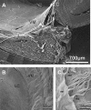
A 75-year-old human eye. (A) Scanning electron micrograph of a sagittal aspect of the anterior vitreous membrane (VM), the posterior tine of the zonular fork (*), and the posterior lens capsule (LC). (B) Scanning electron micrograph of the anterior VM and its attachment to the posterior LC. (C) Careful lifting of the VM reveals that the majority of the zonule (Z) inserts directly into the posterior LC without attachment to the vitreous membrane that itself forms a separate layer also directly inserting into the posterior LC. However, some bundles of zonular fibers (anterior vitreous zonule, aVZ) do insert into the anterior VM at spatial intervals of approximately 75 μm.
Figure 3.
Figure 3.
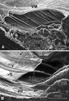
(A) Scanning electron micrographs of a sagittal and oblique internal view of the ciliary body (CB), the vitreous membrane (VM), the zonular plexus (ZP), and the zonular connections bridging the cleft between the pars plana zonules (ppZ) and VM (rhesus monkey, aged 8 years). The bridging bundles of zonular fibers run from the region of the ZP in the valleys of the posterior pars plicata toward the VM in the region of the ora serrata (*). These zonular bundles were termed intermediate vitreous zonule (iVZ). (B) Scanning electron micrograph showing the inner aspect of the posterior pars plicata (ppl), pars plana (pp), VM, and iVZ of a 10-year-old rhesus monkey. Anteriorly, the intermediate vitreous zonular bundles split into a fork with tines that insert on both sides of the processes in the valleys of the ppl (*). Posteriorly, each main bundle splits into several smaller bundles that merge with the VM (arrows).
Figure 4.
Figure 4.
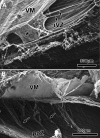
(A) Scanning electron micrographs of sagittal sections of the ora serrata region (rhesus monkey, aged 6 years). The vitreous membrane (VM), the posterior extensions of the intermediate vitreous zonule (iVZ), and the pars plana zonule (ppZ) form an interconnected spongelike structure (*). (B) After careful elevation of the VM, the connections of the ppZ to the posterior VM become visible (arrows).
Figure 5.
Figure 5.
(A) Histologic sagittal section through a 100-year-old human eye showing the posterior adherence of the vitreous membrane to the posterior pars plana and ora region (*). Note that this region in humans encompasses nearly one half the sagittal length between the scleral spur and ora serrata. (B) Scanning electron micrograph of the posterior vitreous zonule (sagittal aspect, 85-year-old human eye). Note the oblique-running fibrils between the pars plana zonules (ppZ) and the vitreous membrane (VM); in contrast to the monkey (Fig. 4B), these fibrils form a latticelike structure (arrows).
Figure 6.
Figure 6.
UBM images of unaccommodated (A) and accommodated (B) ciliary muscle in a live rhesus monkey, aged 25 years. In these somewhat oblique sections, one can see the pars plana zonules immediately adjacent to the pars plana epithelium (arrowheads). This structure is more apparent in the accommodated than in the unaccommodated state. Straight line: the ciliary processes and the ora serrata represent the vitreous zonule. The numbers represent the angle between the anterior face of the ciliary body and the inner surface of the peripheral cornea, as defined by the white lines. Narrowing of the ciliary body (CB)–corneal angle in the accommodated versus the unaccommodated state was used as a surrogate indicator of forward ciliary body movement.
Figure 7.
Figure 7.
UBM images were obtained in a 15-year-old rhesus monkey. Care was taken to assure that the vitreous zonule appeared as a continuous prominent straight line parallel to the focus line (*) imprinted by the instrument within all images, indicating that these were true sagittal sections. (A) Anteriorly, the vitreous membrane is separated from the vitreous zonule. The vitreous zonule fork (seen by SEM, Fig. 3B) occurs just posterior to its insertion to the zonular plexus and, as the fibers split at the fork, they course out of the UBM image plane (arrowhead). Thus, the white line that represents the vitreous zonule appears to discontinue (arrowhead) immediately posterior to reaching the ciliary process region in this image. This drop out of the vitreous zonule strand is not always apparent in UBM images, due to the orientation of the UBM probe and the close proximity of the vitreous zonule fork to the ciliary processes (CP), as in (B).
Figure 8.
Figure 8.
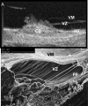
UBM image (A) analogous to SEM sections (B) and (A) of Figure 3 reproduced for ease of comparison, from the same 8-year-old rhesus monkey. CB, ciliary body, pp, pars plana zonule, VZ, vitreous zonule, VM, vitreous membrane.
Figure 9.
Figure 9.
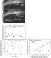
UBM obtained in three rhesus monkeys aged 6, 16, and 25 years, showing the age-related change in cleft width (A–C). Cleft width plotted versus age (D) and accommodative amplitude (E). Note also the decreasing width of the ciliary muscle overlying the widest part of the cleft and the increased curvature of the arc formed by the inner portion of the ciliary body.
Figure 10.
Figure 10.
UBM images in a 21-year-old rhesus monkey eye before (A) and during (B) central electrical supramaximal stimulation of the E-W nucleus. The distance between the scleral spur and the posterior insertion zone of the vitreous zonule was measured in the unaccommodated and accommodated states. CB, ciliary body; c, cornea. (C) Distance between the scleral spur and vitreous zonule insertion zone in the resting eye, and (D) accommodative forward movement of the vitreous zonule insertion zone plotted versus age. (E) Accommodative amplitude plotted versus the forward movement of the vitreous zonule insertion zone during accommodation.
Figure 11.
Figure 11.
Scanning electron micrograph of the temporal region of a 9-year-old rhesus monkey eye after a pars plana injection of α-chymotrypsin shows the absence of intermediate vitreous zonular fibers in this region. VM, vitreous membrane; ppZ, pars plana zonule; CB, ciliary body.
Figure 12.
Figure 12.
Semithin sections of the temporal regions of the right and left eye of a 9-year-old rhesus monkey. In the resting eye (A), the inner edge of the ciliary muscle was located much more anteriorly after α-chymotrypsin injection with disruption of the intermediate vitreous zonule than in the contralateral noninjected eye (B). Sp, scleral spur. Arrow: inner apex.
Figure 13.
Figure 13.
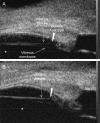
Ultrasound biomicroscopic images of the 25-year-old rhesus monkey eye at rest and during supramaximal central stimulation to induce accommodation. Left: the aphakic eye after ECLE; right: after subsequent α-chymotrypsin lysis of the intermediate vitreous zonule and surgical removal of the lens capsule. The change in the ciliary body to cornea angle during maximal accommodation (bottom) is increased after lysis of the intermediate vitreous zonule fibers (arrows), although the difference at rest (top) is minimal.

References

    1. Lütjen-Drecoll E, Tamm E, Kaufman PL. Age-related loss of morphologic responses to pilocarpine in rhesus monkey ciliary muscle. Arch Ophthalmol 1988;106:1591–1598 - PubMed
    1. Tamm E, Croft MA, Jungkunz W, Lütjen-Drecoll E, Kaufman PL. Age-related loss of ciliary muscle mobility in the rhesus monkey: role of the choroid. Arch Ophthalmol 1992;110:871–876 - PubMed
    1. Croft M, Glasser A, Heatley G, McDonald J, Ebbert T, Kaufman PL. The relationship between accommodative ciliary body and lens function in rhesus monkeys, I: normal lens, zonule and ciliary process configuration in the iridectomized eye. Invest Ophthalmol Vis Sci 2006;47(3):1076–1086 - PubMed
    1. Strenk SA, Semmlow JL, Strenk IM, Munoz P, Gronlund-Jacob J, DeMarco JK. Age-related changes in human ciliary muscle and lens: a magnetic resonance imaging study Invest Ophthalmol Vis Sci 1999;40:1162–1169 - PubMed
    1. Poyer JF, Kaufman PL, Flügel C. Age does not affect contractile response of the isolated monkey ciliary muscle to muscarinic agonists. Curr Eye Res 1993;12:413–422 - PubMed

MeSH terms