. 2009 Oct 9;326(5950):298-301.
doi: 10.1126/science.1175737.

KLF family members regulate intrinsic axon regeneration ability

Affiliations

KLF family members regulate intrinsic axon regeneration ability

Darcie L Moore et al. Science. .

Abstract

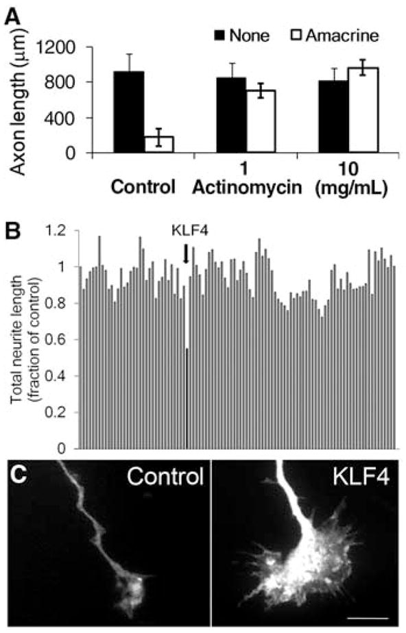
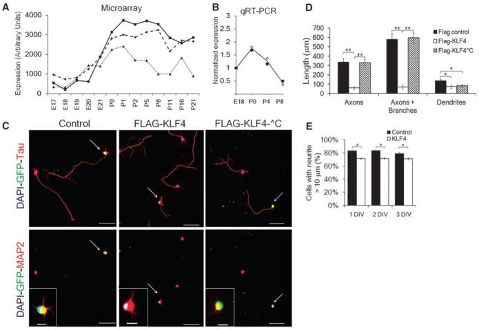
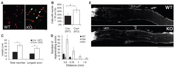
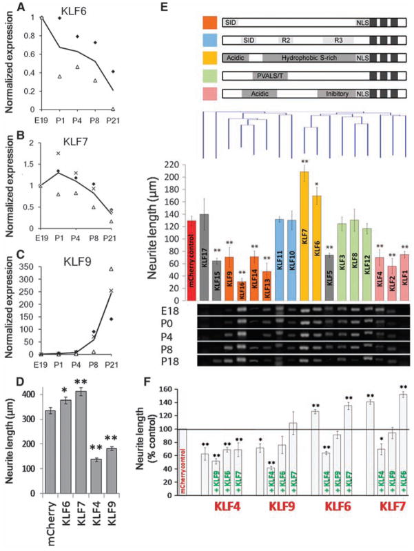
Neurons in the central nervous system (CNS) lose their ability to regenerate early in development, but the underlying mechanisms are unknown. By screening genes developmentally regulated in retinal ganglion cells (RGCs), we identified Krüppel-like factor-4 (KLF4) as a transcriptional repressor of axon growth in RGCs and other CNS neurons. RGCs lacking KLF4 showed increased axon growth both in vitro and after optic nerve injury in vivo. Related KLF family members suppressed or enhanced axon growth to differing extents, and several growth-suppressive KLFs were up-regulated postnatally, whereas growth-enhancing KLFs were down-regulated. Thus, coordinated activities of different KLFs regulate the regenerative capacity of CNS neurons.


Figures

Fig. 1
Fig. 1
A screen of developmentally regulated genes identifies KLF4 as an inhibitor of neurite growth. (A) Purified embryonic RGCs were cultured in the presence (white bars) or absence (black bars) of amacrine cell membranes for 3 days, and replated away from amacrine cell membranes, after which RGC axon growth was measured. Actinomycin D blocked RGCs’ decrease in axon growth caused by amacrine cell membranes (mean ± SEM). (B and C) E18 hippocampal neurons were cotransfected with 111 candidate genes and enhanced green fluorescent protein (EGFP), cultured for 3 days on laminin, and immunostained for Tau to visualize neurites. (B) Neurite length of cotransfected (EGFP+) neurons. Bars represent average neurite length normalized to EGFP control (far left). KLF4 (arrow) decreased neurite growth by 50%. (C) EGFP+ growth cones of EGFP+/KLF4-transfected neurons (right) were enlarged compared to control-transfected neurons (left). Scale bar, 10 μm.
Fig. 2
Fig. 2
KLF4 is developmentally regulated in RGCs, and its overexpression decreases axon growth in a zinc-finger–dependent manner. (A and B) KLF4 expression in RGCs increases at birth, as measured in acutely purified rat RGCs by microarray [three probe sets (A); (19)] or in acutely purified mouse RGCs by qRT-PCR [(B); fold change from E18]. Two biological replicates are plotted with their average in (B). (C to E) FLAG-KLF4-WT, FLAG-KLF4-^C lacking the C-terminal zinc finger DNA binding domain, or FLAG or mCHERRY controls were transfected into E20 RGCs. (C) After 2 days, RGCs were immunostained for FLAG or GFP (green, transfected cells), and Tau or MAP2 (red) as marked [nuclear DAPI (4′,6-diamidino-2-phenylindole) is blue]. Scale bar, 50 μm (main panels), 10 μm (inset). (D) Hand-tracing revealed that FLAG-KLF4-WT overexpression decreased axon growth; overexpression of FLAG-KLF4-^C was similar to that of controls (**P < 0.001, *P < 0.02, unpaired t test, post-Bonferroni correction; mean ± SEM). (E) E20 RGCs were transfected with either mCherry-pIRES2-eGFP (control) or KLF4-pIRES2-EGFP and plated for 1, 2 or 3 days in vitro (DIV). At 1 to 3 DIV, more control-transfected RGCs extended at least one neurite >10 μm than KLF4-transfected RGCs (*P < 0.001, paired t test; mean ± SEM).
Fig. 3
Fig. 3
KLF4 knockout increases RGC neurite growth in vitro and regeneration of adult RGCs in vivo. (A to C) Purified P12 RGCs were cultured from Thy1-cre−/ −/KLF4fl/fl/Rosa+ (Cre− WT) and Thy1-cre+/−/KLF4fl/fl/Rosa+ (Cre+ KO) mice, and plated for 3 DIV before Tau immunostaining and automated tracing. (A) Immunostaining for Tau (red) demonstrated low levels of growth of Cre− WT RGCs (left) but increased levels of axon growth of Cre+ KLF4-KO RGCs (right; Rosa+ yellow cells, arrowheads). (B) KLF4-KO RGCs have a higher percentage of cells with neurites, compared to controls (N = 3; *P < 0.02, t test; mean ± SEM). (C) When all YFP+ RGCs were measured, KLF4 KO RGCs extended longer neurites than WT RGCs (representative experiment shown; *P < 0.001; mean ± SEM). (D and E) Two weeks after optic nerve crush of Thy1-cre+/KLF4+/+ (WT), Thy1-cre+/KLF4fl/+ (Het), and Thy1-cre+/KLF4fl/fl (KO) mice, regenerating fibers were anterogradely labeled by intravitreal injection of Alexa 594–labeled cholera toxin B. Regenerating fibers were counted at specified distances from the lesion site. (D) More fibers regenerate in KO mice compared to WT or Het (n = 10 WT, 4 Het, and 7 KO mice; P < 0.001 for KO versus WT or Het; no difference between WT and Het by mixed-model analysis of covariance; mean ± SEM). (E) Partial projections of sectioned optic nerve from WT and KO mice show regenerating axons more than 1 mm distal to the lesion site in KO nerve. Scale bar, 200 μm.
Fig. 4
Fig. 4
Multiple KLF family members are developmentally regulated in RGCs and differentially affect CNS neurite growth. (A to C) RGCs from multiple ages were purified by immunopanning and analyzed by qRT-PCR. Transcript abundance is normalized to E19. KLF6 (A) and KLF7 (B) decrease more than 5-fold postnatally, whereas KLF9 (C) increases 250-fold. Each marker type is a separate experiment, and the line shown is the average; N = 2 to 3. (D) P4 RGCs were cotransfected with KLFs and EGFP and plated for 2 days on laminin. Bars represent average total neurite length of transfected (EGFP+) neurons [n > 700; *P < 0.05, **P < 0.01; analysis of variance (ANOVA) with post hoc Dunnett’s test; mean ± SEM; pooled data from two replicate experiments]. (E) P5 cortical neurons were cotransfected with individual KLFs and mCherry, plated for 3 days on laminin, and immunostained for β-III tubulin. (Top) KLF family members are grouped according to defined structural domains (27) and clustered by amino acid similarity (Clustal analysis, Vector NTI). (Middle) Bars represent average total neurite length of transfected (mCherry+) neurons and are colored by the presence of known motifs (above). Nine KLFs significantly decreased neurite length, and two increased neurite length (N > 3, n > 100; *P < 0.05, **P < 0.01, ANOVA with post hoc Dunnett’s test; mean ± SEM). (Bottom) Purified RGCs from different ages were analyzed by RT-PCR with KLF-specific primers, ordered according to the overlying bar graph. Transcripts for all KLFs except KLF1 and -17 were detected in developing RGCs. (F) P5 cortical neurons were cotransfected with combinations of KLFs with IRES-mCherry (red) or IRES-EGFP (green) reporters and cultured as above (DNA loading controls, fig. S13). Bars represent average neurite length of dually transfected neurons (mCherry+, EGFP+). Coexpression of KLF4 or -9 blocked the growth-promoting effects of KLF6 or -7 (N = 3, n > 25; *P < 0.05, **P < 0.01, ANOVA with post hoc Dunnett’s test; mean ± SEM).

Comment in

References

    1. Nicholls J, Saunders N. Trends Neurosci. 1996;19:229. - PubMed
    1. Chen DF, Jhaveri S, Schneider GE. Proc Natl Acad Sci USA. 1995;92:7287. - PMC - PubMed
    1. Bregman BS, Kunkel-Bagden E, McAtee M, O’Neill A. J Comp Neurol. 1989;282:355. - PubMed
    1. Yiu G, He Z. Nat Rev Neurosci. 2006;7:617. - PMC - PubMed
    1. Case LC, Tessier-Lavigne M. Curr Biol. 2005;15:R749. - PubMed

Publication types

MeSH terms