. 2009 Nov 4;28(21):3303-14.
doi: 10.1038/emboj.2009.261. Epub 2009 Oct 8.

Amphipathic motifs in BAR domains are essential for membrane curvature sensing

Affiliations

Amphipathic motifs in BAR domains are essential for membrane curvature sensing

Vikram K Bhatia et al. EMBO J. .

Abstract

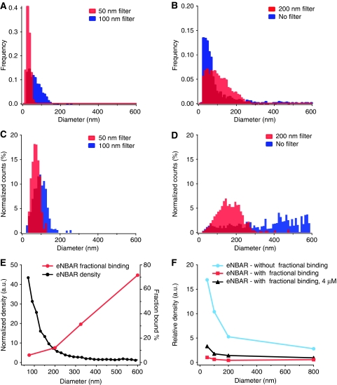
BAR (Bin/Amphiphysin/Rvs) domains and amphipathic alpha-helices (AHs) are believed to be sensors of membrane curvature thus facilitating the assembly of protein complexes on curved membranes. Here, we used quantitative fluorescence microscopy to compare the binding of both motifs on single nanosized liposomes of different diameters and therefore membrane curvature. Characterization of members of the three BAR domain families showed surprisingly that the crescent-shaped BAR dimer with its positively charged concave face is not able to sense membrane curvature. Mutagenesis on BAR domains showed that membrane curvature sensing critically depends on the N-terminal AH and furthermore that BAR domains sense membrane curvature through hydrophobic insertion in lipid packing defects and not through electrostatics. Consequently, amphipathic motifs, such as AHs, that are often associated with BAR domains emerge as an important means for a protein to sense membrane curvature. Measurements on single liposomes allowed us to document heterogeneous binding behaviour within the ensemble and quantify the influence of liposome polydispersity on bulk membrane curvature sensing experiments. The latter results suggest that bulk liposome-binding experiments should be interpreted with great caution.


Conflict of interest statement

The authors declare that they have no conflict of interest.

Figures

Figure 1
Figure 1
BAR domain binding on single liposomes of different diameters and therefore membrane curvature. (A) Sketch of the SLiC assay. Single liposomes are tethered on surfaces using streptavidin–biotin linkage and imaged with confocal microscopy. BAR domains sense membrane curvature by binding with higher density on liposomes of smaller diameter, and this is schematically indicated by the large arrow towards the smaller liposomes. (B, C) Confocal microscopy images of brain liposomes (exc. 633 nm) and BAR protein (eNBAR) (exc. 488 nm), respectively. Scale bar is 3 μm and applies to both images. (D) Intensity profile from cross section of three liposomes indicated by arrows in (B) (274, 95 and 115 nm in diameter calculated by their total intensity), which shows qualitatively membrane curvature sensing by eNBAR. Protein signal (green) is higher for smaller liposomes, notice this effect will be greatly amplified when signals are converted into density. (E) eNBAR density versus liposome diameter for two different concentrations 40 and 400 nM. We typically record a relative eNBAR density increase of ∼20–100-fold. A higher concentration increases the density, i.e. number of proteins pr. unit area. (F) eNBAR normalized density versus liposome diameter for three different concentrations—4, 400 and 6400 nM. We recorded identical membrane curvature sensing graphs for different concentrations. Density is normalized to the baseline (density of largest liposomes was set to 1). (G) A control with the lipid-binding PH domain of Centaurin β2 shows no curvature selectivity as expected. A second control excludes any immobilization artefacts by demonstrating matching membrane curvature sensing curves for liposomes incubated with eNBAR directly in solution (red dots) or after their immobilization on a surface (black dots). Protein concentration was 40 nM.
Figure 2
Figure 2
Comparison of membrane curvature sensing graphs for various BAR domains. Testing the current hypothesis of the molecular mechanism of membrane curvature sensing by BAR domains. All samples are incubated with protein at 40 nM for 1 h unless otherwise stated. (A) Representative members of three BAR domain super families. Surface representation coloured according to electrostatic potential and mesh equipotential surfaces at −5 kT/e to 5 kT/e (blue is +, red is −). Arrows indicate the position of AHs. Neither dimer shape nor electrostatic potential is conserved for the different proteins. (B) Comparison of eNBAR and eNBAR-3KE shows that charge mutations do not alter the ability of this BAR domain to sense membrane curvature. Sensing is identical for eNBAR 3KE at high concentration (5.2 μM). (C) Comparison of eNBAR and oFBAR shows that the intrinsic radius of curvature of the dimer (11 nm versus 65 nm) does not influence membrane curvature sensing. (D) eNBAR shows identical membrane curvature sensing as an IBAR (mIBAR), which has an negative radius of curvature according to the crystal structure. Sensing is identical for mIBAR at high concentration (0.9 μM). Thus, neither charges nor BAR dimer structure is essential for curvature sensing.
Figure 3
Figure 3
Binding curves of eNBAR on membranes of different curvature. (A) Quantitative determination of the concentration-dependent binding of eNBAR as a function of liposome diameter. Depicted are representative binding curves for three different liposome diameters fitted with Langmuir isotherms (100, 200 and 650 nm). Strikingly, the eNBAR shows a strong curvature selectivity even at saturation. (B) From the fits in (A) we extract the size dependency of the apparent equilibrium disassociation constant (KD) and the apparent free energy of binding. The KD varies from ∼400 to 140 nM illustrating a marginally higher affinity of eNBAR for curved membranes. (C) The saturation density (Bmax) extracted from (A) as a function of liposome diameter. The size dependence of the Bmax changes to a much greater extent than KD. The inset depicts the Bmax in a log–log plot. Membrane curvature sensing is not mediated by higher affinity but relies predominantly on a curvature-dependent Bmax.
Figure 4
Figure 4
Membrane curvature sensing of BAR domains is dominated by the N-terminal amphipathic helix. All graphs are measured at 40 nM protein unless otherwise stated. (A) Membrane curvature sensing by eN fully replicates the sensing ability of eNBAR. (B) Helical wheel representation of eN, arrow indicates the point mutation F10E. Yellow, indicates hydrophobic residues; green, polar; blue, basic; pink, acidic. (C) Membrane curvature sensing graph for eNBAR-F10E measured at 740 nM. The point mutation disrupting the formation of the N-terminal AH impaired severe membrane curvature sensing. The concentration was increased to get sufficient binding. (D) The data for aN and aNBAR overlay completely with the fit for eNBAR showing the three molecules have identical membrane curvature sensing ability. (E) Helical wheel representation of aN, arrows indicate point mutations to glutamate (3xE). (F) Membrane curvature sensing of aNBAR was also severely impaired by mutations in the N-terminal peptide (aNBAR 3xE), binding measured at 4 μM. Thus, membrane curvature sensing by BAR domains seems to originate from the insertion of the N-terminal AHs in the bilayer.
Figure 5
Figure 5
Membrane curvature sensing by BAR domains is inhibited by blocking the curvature-induced hydrophobic defects of a bilayer. Graphs are measured at 40 nM protein unless otherwise stated. (A) Sketch showing blockage of membrane curvature-induced defects in the bilayer by pre-incubation of liposomes with a small amphiphilic molecule (C18-fluorescein). (B) Blocking of defects impaired membrane curvature sensing mediated by hydrophobic insertion for an AH (eN). Inset: typical line scan showing qualitatively that eN binds to blocked liposomes but without discriminating different diameters. (C) Control with PH domain (1 μM) on normal and blocked liposomes. Both conditions show identical absolute density assuring that blocking with amphiphiles does not cause any general lipid-binding effects. (DF) Blocking of defects impaired membrane curvature sensing for eNBAR, aNBAR and mIBAR, respectively at both high (2, 1 and 0.9 μM, respectively) and low concentration, further supporting that membrane curvature sensing by BAR domains depends on curvature-induced defects in the bilayer and not charge-mediated interactions. (G) Control with eNBAR F10E at 5 μM also show that the residual binding from a BAR domain is not capable of sensing through charge-mediated interactions.
Figure 6
Figure 6
Heterogeneous binding of eNBAR wild type and mutants showed in single liposome measurements. All graphs are measured at 40 nM protein unless otherwise stated. (A) Typical binding kinetics for eNBAR to four single liposomes (blue, red, black and green traces). A variable lag phase is identified lasting up to several tens of minutes. (B) Heterogeneous binding kinetics results in a variable fraction of liposomes showing binding (Bfrac). Bfrac is documented for different binding conditions (protein incubation (1 h) in solution or on the surface) and several different binding partners (eNBAR, PH domain (1 μM) and eN). (C) Measured eNBAR Bfrac as a function of liposome diameter for two different concentrations within a fixed incubation time (1 h). Error bars represent the s.d. for three different samples of approximately 800–1000 liposomes each. (D) Bfrac for different eNBAR and eN mutants. Bfrac is primarily affected by a point mutation in the AH (F10E) and to a lesser degree by mutations that modify the electrostatic potential on the concave side of eNBAR (3KE). Incubation time and error bars as in (C). A full-colour version of this figure is available at The EMBO Journal Online.
Figure 7
Figure 7
Reconstruction of bulk membrane curvature measurements from single liposome data. (A, B) Distribution of liposome diameters for populations extruded with filters of different pore size (50, 100 and 200 nm) or not extruded at all. (C, D) Distribution of the lipid mass at given diameter range. Conditions as in (A, B). (E) eNBAR density (black) and fractional binding (red) measured at 40 nM. (F) Simulation of bulk ensemble averaging without fractional binding (Bfrac) (blue), with Bfrac at 40 nM (red) or 4000 nM (black). Bulk ensemble assays may potentially provide false-negative results because of a convolution of density and fractional binding. A full-colour version of this figure is available at The EMBO Journal Online.

References

    1. Bagatolli LA (2006) To see or not to see: lateral organization of biological membranes and fluorescence microscopy. Biochim Biophys Acta Biomembr 1758: 1541–1556 - PubMed
    1. Bendix PM, Pedersen MS, Stamou D (2009) Quantification of nano-scale intermembrane contact areas by using fluorescence resonance energy transfer. Proc Natl Acad Sci USA 106: 12341–12346 - PMC - PubMed
    1. Bigay J, Casella JF, Drin G, Mesmin B, Antonny B (2005) ArfGAP1 responds to membrane curvature through the folding of a lipid packing sensor motif. EMBO J 24: 2244–2253 - PMC - PubMed
    1. Blood PD, Swenson RD, Voth GA (2008) Factors influencing local membrane curvature induction by N-BAR domains as revealed by molecular dynamics simulations. Biophys J 95: 1866–1876 - PMC - PubMed
    1. Blood PD, Voth GA (2006) Direct observation of Bin/amphiphysin/Rvs (BAR) domain-induced membrane curvature by means of molecular dynamics simulations. Proc Natl Acad Sci USA 103: 15068–15072 - PMC - PubMed

Publication types

MeSH terms