. 2009 Oct;5(10):e1000611.
doi: 10.1371/journal.ppat.1000611. Epub 2009 Oct 9.

A granulin-like growth factor secreted by the carcinogenic liver fluke, Opisthorchis viverrini, promotes proliferation of host cells

Affiliations

A granulin-like growth factor secreted by the carcinogenic liver fluke, Opisthorchis viverrini, promotes proliferation of host cells

Michael J Smout et al. PLoS Pathog. 2009 Oct.

Abstract

The human liver fluke, Opisthorchis viverrini, infects millions of people throughout south-east Asia and is a major cause of cholangiocarcinoma, or cancer of the bile ducts. The mechanisms by which chronic infection with O. viverrini results in cholangiocarcinogenesis are multi-factorial, but one such mechanism is the secretion of parasite proteins with mitogenic properties into the bile ducts, driving cell proliferation and creating a tumorigenic environment. Using a proteomic approach, we identified a homologue of human granulin, a potent growth factor involved in cell proliferation and wound healing, in the excretory/secretory (ES) products of the parasite. O. viverrini granulin, termed Ov-GRN-1, was expressed in most parasite tissues, particularly the gut and tegument. Furthermore, Ov-GRN-1 was detected in situ on the surface of biliary epithelial cells of hamsters experimentally infected with O. viverrini. Recombinant Ov-GRN-1 was expressed in E. coli and refolded from inclusion bodies. Refolded protein stimulated proliferation of murine fibroblasts at nanomolar concentrations, and proliferation was inhibited by the MAPK kinase inhibitor, U0126. Antibodies raised to recombinant Ov-GRN-1 inhibited the ability of O. viverrini ES products to induce proliferation of murine fibroblasts and a human cholangiocarcinoma cell line in vitro, indicating that Ov-GRN-1 is the major growth factor present in O. viverrini ES products. This is the first report of a secreted growth factor from a parasitic worm that induces proliferation of host cells, and supports a role for this fluke protein in establishment of a tumorigenic environment that may ultimately manifest as cholangiocarcinoma.


Conflict of interest statement

The authors have declared that no competing interests exist.

Figures

Figure 1
Figure 1. Comparison of multiple-reaction monitoring (MRM) MS/MS fragmentation spectra targeted against a 19 amino acid Ov-GRN-1 peptide identified during the course of shotgun proteomics.
Recombinant Ov-GRN-1 (red) and O. viverrini ES products (blue) were analyzed in separate MRM experiments targeted against a previously identified Ov-GRN-1 peptide. In both experiments the target precursor ion eluted from the reverse-phase column at the same time and produced identical MS/MS fragmentation patterns. The MS/MS spectrum from the original identification is shown in the inset and the identified fragments are labeled. Peaks corresponding to the MS/MS spectra are highlighted with an asterisk on the MRM fragmentation spectrum along with the triple charged precursor ions.
Figure 2
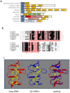
Figure 2. Ov-GRN-1 is a secreted growth factor belonging to the granulin family.
Panel A - structural architecture of proteins containing granulin domains. Grn or GrnA-G: granulin domains, Inhib: cathepsin propeptide inhibitor, FN: Fibronectin type III, Sig: Secretion Signal sequence, Protease: Papain family cysteine protease, Esterase: SGNH hydrolase type esterase. Sequence accession numbers are as described in the legend for Figure 2, with structural architecture obtained from Interpro 18.0 database. Panel B - multiple sequence alignment of Ov-GRN-1 and granulin domains from other organisms. Identical amino acids are in black boxes and similar amino acids are in gray boxes. The granulin core domain is highlighted by a pink box. Theoretical disulphide bonds are numbered one to six above each cysteine residue. GenBank accession numbers are as follows: Opisthorchis viverrini Ov-GRN-1- FJ436341*; Clonorchis sinensis (parasitic liver fluke) - AT006891*; Schistosoma japonicum (parasitic blood fluke) - AY810079*; Caenorhabditis elegans (free living roundworm) - NP_492982*; Homo sapiens (human) granulin domains A and B - AW51601; Mus musculus (mouse) - NM_008175*. *Amino acid numbering is from the initiator (or predicted initiator) methionine. Panel C - molecular model of the granulin core domain of Ov-GRN-1 based on the nuclear magnetic resonance structure of carp granulin and an overlay where Ov-GRN-1 is superimposed on carp granulin. Disulphide bonds are highlighted in yellow.
Figure 3
Figure 3. Neighbor joining phylogenetic tree for the granulin core domains from a range of phyla.
Numbers on the branches are bootstrap values that obtained greater than 50% support from 1000 replicates. GenBank accession numbers are as follows: Ov-GRN-1 - FJ436341; Cs- Clonorchis sinensis (parasitic liver fluke) AT006891, Sj - Schistosoma japonicum (parasitic blood fluke) AAX25968; Mx - Myxococcus xanthus (bacterium) Q1CVL1; Nc - Neurospora crassa (fungus) Q7S2H4; Dr - Danio rerio (zebrafish) A8E5C4; Xt - Xenopus tropicalis (frogs) CAJ82256; Nv - Nematostella vectensis (sea anemone) A7SM19; Aa - Aedes albopictus (mosquito) B0FU00; Os - Oryza sativa (rice) Q7XR52; Eg - Elaeis guineensis var. tenera (oil palm) A6N8F8; Ta - Trichoplax adhaerens (tablet animals) B3RQV7; Dd - Dictyostelium discoideum (amoeba) Q54QR7; At - Arabidopsis thaliana (mouse-ear cress) Q9LT78; Ce - Caenorhabditis elegans (free living roundworm) BAE35565; Hs - Homo sapiens (human) EAW51601; Mm - Mus musculus (mouse) NP_492982.
Figure 4
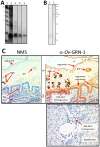
Figure 4. Recombinant Ov-GRN-1e expressed in E. coli and verification of anti-Ov-GRN-1 antibodies.
Panel A: 15% SDS PAGE gel stained with Imperial Blue showing purification of Ov-GRN-1e. Lane 1: Benchmark Marker (Invitrogen), 2: French pressed total cell pellet, 3: Insoluble fraction, 4: column eluate containing purified recombinant Ov-GRN-1e, 5: refolded Ov-GRN-1e. Panel B: Recognition of native Ov-GRN-1 in O. viverrini somatic protein extract by mouse anti-Ov-GRN-1s IgG. SDS-PAGE was conducted under native conditions and the blot was probed with control IgG (1) or anti-Ov-GRN-1s IgG (2). The arrow highlights the major band at 38 kDa. Molecular masses (kDa) are indicated on the right. Panel C: Immunolocalization of Ov-GRN-1 in histological sections of adult O. viverrini in the bile ducts of experimentally infected hamsters. The left panel was probed with control IgG; the right panel was probed with anti-Ov-GRN-1e IgG. The bottom panel shows a liver section from an uninfected hamster that was probed with anti-Ov-GRN-1e IgG. Peroxidase staining is revealed as a brown/rust colored deposit in panel B and Mayer's Haematoxylin counterstained the nuclei in blue. Red arrows highlight the regions within in the O. viverrini parasite and bile duct tissue that stained positive for Ov-GRN-1.
Figure 5
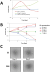
Figure 5. Proliferation of host cells in response to O. viverrini proteins.
Panel A: Non-contact co-culture of 3 or 10 living adult O. viverrini worms with KKU100 human biliary cancer cell line over time. Growth is expressed as a ratio of cells cultured in the presence or absence of O. viverrini. Cell numbers were manually counted using a hemocytometer. Panel B: Effect of different concentrations of O. viverrini adult worm ES on the growth of NIH-3T3 mouse fibroblasts over time. Cell growth was measured using WST-1 metabolic assay as a marker of mitochondrial enzyme activity. Growth is expressed as a ratio compared to cells that did not receive ES with error bars indicating one standard deviation. Panel C: Morphology changes of NIH-3T3 mouse fibroblasts cultured in the presence or absence of O. viverrini adult worm ES (20 µg/ml) after 3 days.
Figure 6
Figure 6. Recombinant refolded Ov-GRN-1e stimulates growth of NIH-3T3 fibroblasts at nanomolar concentrations as measured by WST-1 cell count assay.
Growth is expressed as a ratio of cell numbers after incubation with recombinant proteins compared to PBS (growth rate = 1.0). Panel A: The effects of Ov-GRN-1e and control protein on NIH 3T3 cells for 3 days. To facilitate view of error bars, data points were offset by 0.05 units. Panel B: Growth of cells after three days of culture in the presence of 2–400 nM Ov-GRN-1e and Sm-TSP-2 control protein. All curves are the averages of duplicates and standard deviations are shown as error bars at each data point. Asterisks denote significance at the P<0.05 (*), P<0.01 (**) and P<0.001 (***) levels between growth ratios of cells cultured with Ov-GRN-1e and negative control protein at equivalent concentrations.
Figure 7
Figure 7. Refolded recombinant Ov-GRN-1e stimulates growth of NIH-3T3 fibroblasts at nanomolar concentrations as measured in real time using an xCELLigence system (Roche).
The curves represent the difference between control cells grown in medium plus PBS versus test cells (recombinant proteins added) as measured by Cell Index; data have been normalized prior to sample addition. The control protein and Ov-GRN-1e were added to cells at equimolar quantities, as previously determined and optimized using the WST-1 assay. Ov-GRN-1e before and after (column flow-through) filtration through a 3 kDa cut-off nanosep filter are shown.
Figure 8
Figure 8. Inhibition of ES-induced cell proliferation by anti-Ov-GRN-1 IgG.
Panel A: Anti-Ov-GRN-1s IgG inhibited growth in a dose-dependent manner of NIH-3T3 fibroblasts induced by O. viverrini ES products as measured by the WST-1 metabolic assay. Panel B: Anti-Ov-GRN-1e and anti-Ov-GRN-1s IgGs inhibited growth of NIH-3T3 fibroblasts induced by O. viverrini ES as measured in real time using an xCELLigence system (Roche). Panel C: Anti-Ov-GRN-1e and anti-Ov-GRN-1s IgGs inhibited growth of KKU-100 cholangiocarcinoma cells induced by O. viverrini ES in a dose-dependent manner. Data were obtained using the WST-1 metabolic assay. All data shown are means of triplicate experiments and standard deviations at each data point are shown as error bars. Asterisks denote significance at the P<0.05 (*), P<0.01 (**) and P<0.001 (***) levels between growth ratios or absorbance readings of cells cultured with anti-Ov-GRN-1 and negative control antibodies at equivalent bovine calf serum concentrations.

References

    1. Sripa B, Pairojkul C. Cholangiocarcinoma: lessons from Thailand. Curr Opin Gastroenterol. 2008;24:349–356. - PMC - PubMed
    1. Parkin DM. The global health burden of infection-associated cancers in the year 2002. Int J Cancer. 2006;118:3030–3044. - PubMed
    1. Herrera LA, Benitez-Bribiesca L, Mohar A, Ostrosky-Wegman P. Role of infectious diseases in human carcinogenesis. Environ Mol Mutagen. 2005;45:284–303. - PubMed
    1. Thuwajit C, Thuwajit P, Kaewkes S, Sripa B, Uchida K, et al. Increased cell proliferation of mouse fibroblast NIH-3T3 in vitro induced by excretory/secretory product(s) from Opisthorchis viverrini. Parasitology. 2004;129:455–464. - PubMed
    1. Sripa B, Kaewkes S, Sithithaworn P, Mairiang E, Laha T, et al. Liver Fluke Induces Cholangiocarcinoma. PLoS Med. 2007;4:e201. doi: 10.1371/journal.pmed.0040201. - DOI - PMC - PubMed

Publication types

MeSH terms