. 2009 Oct;5(10):e1000619.
doi: 10.1371/journal.ppat.1000619. Epub 2009 Oct 9.

Virus-Induced Chaperone-Enriched (VICE) domains function as nuclear protein quality control centers during HSV-1 infection

Affiliations

Virus-Induced Chaperone-Enriched (VICE) domains function as nuclear protein quality control centers during HSV-1 infection

Christine M Livingston et al. PLoS Pathog. 2009 Oct.

Abstract

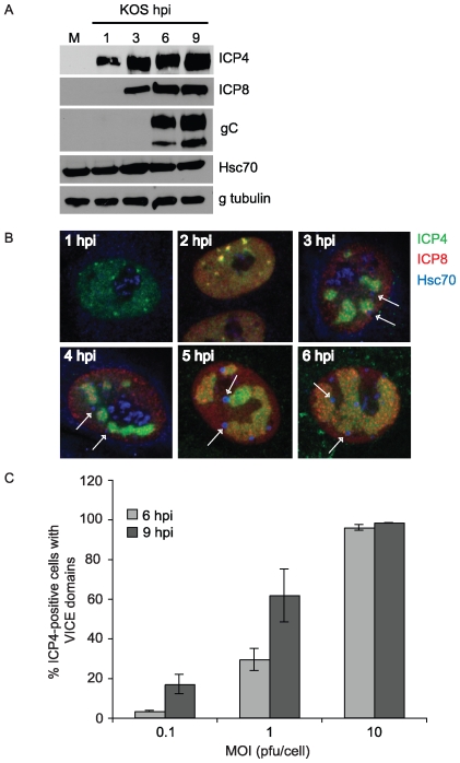
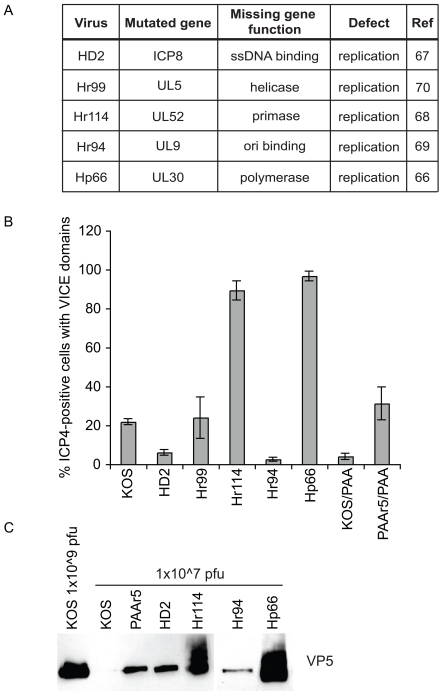
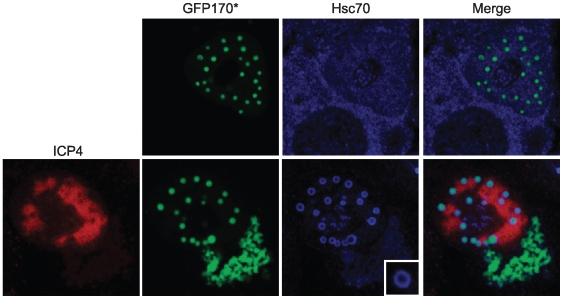
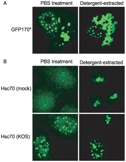
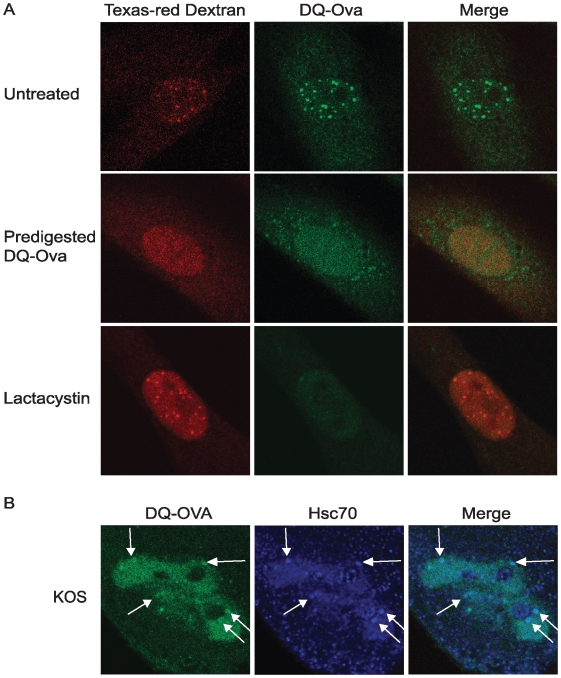
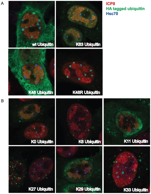
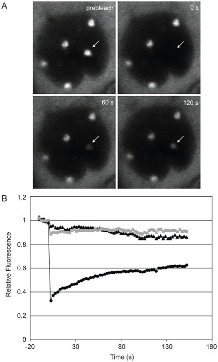
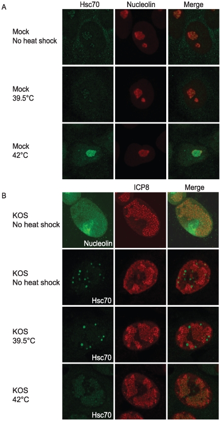
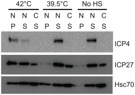
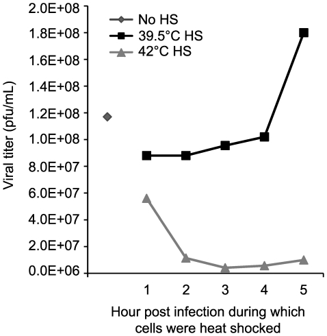
Virus-Induced Chaperone-Enriched (VICE) domains form adjacent to nuclear viral replication compartments (RC) during the early stages of HSV-1 infection. Between 2 and 3 hours post infection at a MOI of 10, host protein quality control machinery such as molecular chaperones (e.g. Hsc70), the 20S proteasome and ubiquitin are reorganized from a diffuse nuclear distribution pattern to sequestration in VICE domains. The observation that VICE domains contain putative misfolded proteins suggests that they may be similar to nuclear inclusion bodies that form under conditions in which the protein quality control machinery is overwhelmed by the presence of misfolded proteins. The detection of Hsc70 in VICE domains, but not in nuclear inclusion bodies, indicates that Hsc70 is specifically reorganized by HSV-1 infection. We hypothesize that HSV-1 infection induces the formation of nuclear protein quality control centers to remodel or degrade aberrant nuclear proteins that would otherwise interfere with productive infection. Detection of proteolytic activity in VICE domains suggests that substrates may be degraded by the 20S proteasome in VICE domains. FRAP analysis reveals that GFP-Hsc70 is dynamically associated with VICE domains, suggesting a role for Hsc70 in scanning the infected nucleus for misfolded proteins. During 42 degrees C heat shock, Hsc70 is redistributed from VICE domains into RC perhaps to remodel viral replication and regulatory proteins that have become insoluble in these compartments. The experiments presented in this paper suggest that VICE domains are nuclear protein quality control centers that are modified by HSV-1 to promote productive infection.


Conflict of interest statement

The authors have declared that no competing interests exist.

Figures

Figure 1
Figure 1. VICE domain formation increases with MOI and duration of infection.
In panel A, Vero cells were mock-infected or infected with KOS at an MOI of 10 for 1, 3, 6 and 9 hours. Samples were harvested in 2× DTT-containing SDS sample buffer and resolved on a 10% SDS-PAGE gel. PVDF membranes were reacted with mouse-anti-ICP4, mouse-anti-ICP8, mouse-anti-glycoprotein C (gC), rat-anti-Hsc70 and mouse-anti-γ tubulin antibodies. In panel B, Vero cells were infected with KOS at an MOI of 10 for 1, 2, 3, 4, 5 and 6 hours followed by fixation and permeabilization in preparation for IF analysis. Samples were triple-labeled with mouse-anti-ICP4, rabbit-anti-ICP8 367 and rat-anti-Hsc70 antibodies. Arrows point to VICE domains that formed adjacent to developing RCs. Images were obtained at 63× magnification with 2× digital zoom. In panel C, Vero cells adhered to glass coverslips were infected with KOS at MOIs 0.1, 1, and 10 for 6 and 9 hours. Cells were fixed, permeabilized and double-labeled with mouse-anti-ICP4 and rat-anti-Hsc70 antibodies. At least 100 ICP4-positive cells of each sample were assessed for whether or not they contained VICE domains (Hsc70 foci). ICP4-negative cells never contained Hsc70 foci. The data represent at least 3 independent experiments and error bars were calculated from standard error of the mean.
Figure 2
Figure 2. Viral stocks with high particle-to-pfu ratio induce the formation of VICE domains.
The replication mutant viruses used in this study are detailed in panel A. In panel B, Vero cells were infected with KOS, HD2 (ICP8 mutant), Hr99 (UL5 mutant), Hr114 (UL52 mutant), Hr94 (UL9 mutant), Hp66 (UL30 mutant) or PAAr5 (PAA-resistant KOS) for 6 hours at an MOI of 2 in the presence or absence of 400 ug/mL PAA (where indicated). At least 100 ICP4-positive cells of each sample were assessed for whether or not they had formed VICE domains. The data represent 3 independent experiments and error bars were calculated from standard error of the mean. In panel C, Viral stocks were normalized to 1e7 pfu/mL based on known titers and loaded directly to an 8% SDS-PAGE gel. Proteins were transferred to a PVDF membrane and blotted for the major capsid protein VP5.
Figure 3
Figure 3. A model misfolded protein is localized to VICE domains.
Vero cells were transfected with 1 ug of plasmid DNA encoding the model misfolded protein GFP170* . Following about 16 hours of protein expression, the cells were infected with KOS at an MOI of 10 for 6 hours. Cells were fixed, permeabilized and labeled with rabbit-anti-ICP8 367 and rat-anti-Hsc70 antibodies. The inset in the bottom row shows an enlarged image of a VICE domain containing GFP170*.
Figure 4
Figure 4. VICE domains are resistant to detergent extraction.
(A) Vero cells were transfected with 1 ug GFP170* . Following 16 hours of protein expression, cells were treated with PBS (left panel) or 0.5% TX-100 detergent extraction buffer (right panel) for 2 minutes on ice. Cells were then fixed in 4% paraformaldehyde, permeabilized in 1% TX-100 and analyzed for localization of GFP170*. (B) Vero cells were mock infected or infected with KOS at an MOI of 10. Following 6 hours of infection, cells were treated with PBS or detergent extraction buffer as described in (A). Hsc70 localization was detected with a rat-anti-Hsc70 (Stressgen) antibody. Imaging was performed on a Zeiss LSM 410 confocal microscope. The gain was set to visualize the diffuse Hsc70, resulting in saturation of the VICE domains.
Figure 5
Figure 5. Protease activity is detected in VICE domains.
(A) HFF-1 cells were plated in dishes containing embedded glass coverslips and were co-microinjected with TX red dextran and the 20S proteasomal substrate DQ-OVA. Images of live cells are shown. In the middle row, cells were co-microinjected with TX red dextran and DQ-OVA that was predigested in the presence of trypsin. In the bottom row, cells were co-microinjected with TX red dextran, DQ-OVA and lactacystin. (B) HFF-1 cells plated in dishes with embedded coverslips were infected with 2 pfu/cell KOS and microinjected 5 hours later with TX red dextran and DQ-OVA. Following about 20–30 min of proteolytic cleavage, the cells were fixed, permeabilized and stained for Hsc70.
Figure 6
Figure 6. Homotypic ubiquitin chains are not detected in VICE domains.
Vero cells adhered to glass coverslips were transfected with 1 ug of pRK-HA-Ub constructs encoding wt, K63, K48 and K48R Ub (panel A) and K0 (monomeric), K6, K11, K27, K29 and K33 Ub (panel B). Following 16–18 hours of protein expression, transfected cells were infected with KOS at an MOI of 10 for 6 hours. At harvest, cells were prepared for IF analysis and labeled with mouse-anti-HA tag, rabbit-anti-ICP8 367 and rat-anti-Hsc70 antibodies. Images were taken at 63× magnification with 2× digital zoom. Merged images of green, red and far-red channels are shown.
Figure 7
Figure 7. FRAP analysis of Hsc70-GFP in VICE domains.
Vero cells adhered to a coverslip dish were transfected with 1 ug of a plasmid encoding wt Hsc70-GFP. Approximately 16 hours post transfection, cells were infected with KOS at an MOI of 10. At 5 hours post infection, live infected cells were imaged on the LSM 510 Meta confocal microscope. At time 0, Hsc70-GFP in one VICE domain was photobleached using 100% argon laser power, and subsequent images collected at ∼2.5 s intervals. (A) Single images from the time series are shown; arrow indicates the VICE domain selected for photobleaching. (B) Fluorescence intensity in the bleached VICE domain (circles), a VICE domain that was not photobleached (squares) and an equivalently sized area of the nucleoplasm (triangles) is shown. Data are normalized to prebleach intensity and corrected for bleaching during monitoring. The experiment shown is representative of at least three independent experiments each involving analysis of numerous infected cells.
Figure 8
Figure 8. Hsc70 localizes to replication compartments in 42°°°°°°C heat-shocked cells.
In panel A, mock infected cells were left at 37°C or heat shocked at 42° or 39.5°C for 1 hour followed by harvest for IF analysis of Hsc70 localization. In panel B, Vero cells were infected with KOS at an MOI of 10. Top row: infected cells were double labeled for nucleolin and ICP8. Bottom three rows: At 5 hours post infection, cells were either left at 37°C or transferred to a 42°C incubator for 1 hour. All samples were harvested at 6 hours post infection and were double labeled for Hsc70 and ICP8.
Figure 9
Figure 9. Viral proteins lose solubility during 42°°°°°°C heat shock.
Vero cells were infected with 10 pfu/cell KOS for 6 hours. During the 5th hour of infection, samples were either left at 37°C or heat shocked for 1 hour at 42°C or 39.5°C. Whole cell extract was fractionated into nuclear (N) soluble (S), nuclear insoluble (P) and cytosolic (C) fractions as described in Materials and Methods, resolved on 10% SDS-PAGE gels and transferred to PVDF membrane. Membranes were probed with monoclonal antibodies specific for ICP4, ICP27 or Hsc70.
Figure 10
Figure 10. Viral productivity decreases following heat shock.
Vero cells were infected with 10 pfu/cell and heat shocked at either 39.5°C or 42°C during the indicated hour post infection before shift back to 37°C. Cell-associated and supernatant virus was harvested 24 hours post infection followed by titration on Vero cell monolayers in triplicate.

References

    1. Outeiro TF, Tetzlaff J. Mechanisms of disease II: cellular protein quality control. Semin Pediatr Neurol. 2007;14:15–25. - PubMed
    1. Fu L, Gao YS, Tousson A, Shah A, Chen TL, et al. Nuclear aggresomes form by fusion of PML-associated aggregates. Mol Biol Cell. 2005;16:4905–4917. - PMC - PubMed
    1. Matsumoto G, Kim S, Morimoto RI. Huntingtin and mutant SOD1 form aggregate structures with distinct molecular properties in human cells. J Biol Chem. 2006;281:4477–4485. - PubMed
    1. Paulson HL, Perez MK, Trottier Y, Trojanowski JQ, Subramony SH, et al. Intranuclear inclusions of expanded polyglutamine protein in spinocerebellar ataxia type 3. Neuron. 1997;19:333–344. - PubMed
    1. Stenoien DL, Mielke M, Mancini MA. Intranuclear ataxin1 inclusions contain both fast- and slow-exchanging components. Nat Cell Biol. 2002;4:806–810. - PubMed

Publication types