. 2010 May 15;126(10):2319-29.
doi: 10.1002/ijc.24920.

Role of CXC chemokine ligand 13 in oral squamous cell carcinoma associated osteolysis in athymic mice

Affiliations

Role of CXC chemokine ligand 13 in oral squamous cell carcinoma associated osteolysis in athymic mice

Subramanya N M Pandruvada et al. Int J Cancer. .

Abstract

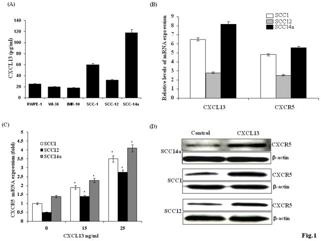
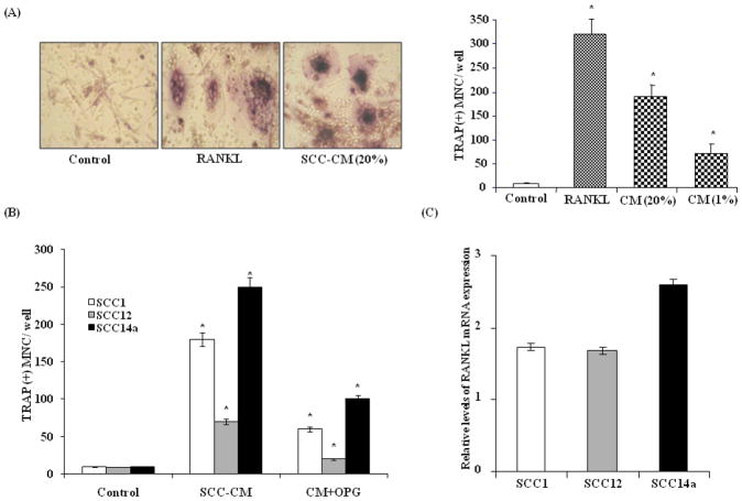
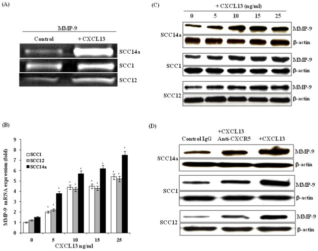
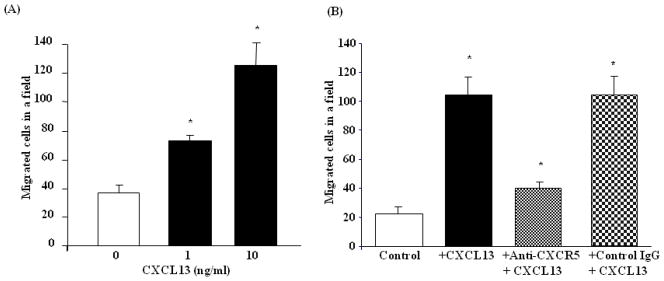
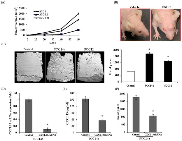
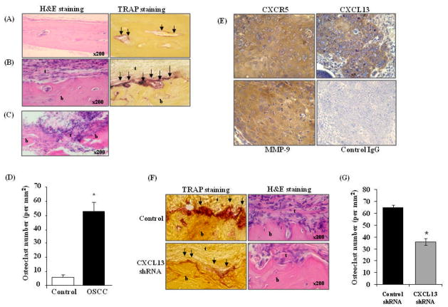
Oral squamous cell carcinomas (OSCC) are malignant tumors with a potent activity of local bone invasion; however, the molecular mechanisms of tumor osteolysis are unclear. In this study, we identified high level expression of chemokine ligand, CXCL13 and RANK ligand (RANKL) in OSCC cells (SCC1, SCC12 and SCC14a). OSCC cell-conditioned media (20%) induced osteoclast differentiation which was inhibited by OPG in peripheral blood monocyte cultures indicating that OSCC cells produce soluble RANKL. Recombinant hCXCL13 (10 ng/ml) significantly enhanced RANKL-stimulated osteoclast differentiation in these cultures. Trans-well migration assay identified that CXCL13 induces chemotaxis of peripheral blood monocytes in vitro which was inhibited by addition of anti-CXCR5 receptor antibody. Zymogram analysis of conditioned media from OSCC cells revealed matrix metalloproteinase-9 (MMP-9) activity. Interestingly, CXCL13 treatment to OSCC cells induced CXCR5 and MMP-9 expression suggesting an autocrine regulatory function in OSCC cells. To examine the OSCC tumor cell bone invasion/osteolysis, we established an in vivo model for OSCC by subcutaneous injection of OSCC cells onto the surface of calvaria in NCr-nu/nu athymic mice, which developed tumors in 4-5 weeks. muCT analysis revealed numerous osteolytic lesions in calvaria from OSCC tumor-bearing mice. Histochemical staining of calvarial sections from these mice revealed a significant increase in the numbers of TRAP-positive osteoclasts at the tumor-bone interface. Immunohistochemical analysis confirmed CXCL13 and MMP-9 expression in tumor cells. Thus, our data implicate a functional role for CXCL13 in bone invasion and may be a potential therapeutic target to prevent osteolysis associated with OSCC tumors in vivo.


Figures

Figure 1
Figure 1
CXCL13/CXCR5 expression in human OSCC cells. (A). CXCL13 levels in conditioned media (CM) obtained from OSCC cells (SCC14a, SCC12, SCC1), normal epithelial cells (RWPE-1) and normal fibroblast cells (WI-38 and IMR-90) as measured by ELISA. (B). Real-time RT-PCR analysis of CXCL13 and CXCR5 receptor expression relative to the level of GAPDH amplification in OSCC cells. (C) CXCL13 stimulates CXCR5 mRNA expression in OSCC cells. The cells were stimulated with different concentrations of CXCL13 (0–25 ng/ml) for 48 h. Total RNA was isolated from these cells and CXCR5 mRNA expression was quantified by Real time RT-PCR analysis (*p<0.05). (D) Western blot analysis of CXCR5 expression in CXCL13 stimulated OSCC cells.
Figure 2
Figure 2
Conditioned media (CM) from OSCC cells stimulates osteoclastogenesis. (A) SCC14a cell CM induced osteoclast differentiation in human peripheral blood monocyte (PBMC) culture. PBMC were incubated with RANKL (100 ng/ml) or SCC-CM (1 and 20%) in the presence of M-CSF (10 ng/ml) for 10 days. Cells cultured with M-CSF alone served as control. TRAP-positive multinucleated osteoclasts formed at the end of the culture period were scored (*p<0.05). (B) Osteoprotegerin (OPG) inhibition of OSCC-CM stimulated osteoclast differentiation in PBMC cultures. PBMC cultured with OSCC-CM (20%) with and without OPG (100 ng/ml) for 10 days. PBMC cultured with M-CSF alone served as control. TRAP positive multinucleated cells formed at the end of the culture period were scored (*p<0.05). (C) Real-time RT-PCR analysis of RANKL mRNA expression in OSCC cells. Relative mRNA expression level was normalized with respect to GAPDH amplification (*p<0.05).
Figure 3
Figure 3
CXCL13 stimulates MMP-9 expression in OSCC cells. (A) Gelatin zymogram analysis of MMP-9 activity in CM obtained from OSCC cells (SCC14a, SCC1 and SCC12) were stimulated with and without CXCL13 (10 ng/ml) for 48 h. (B) OSCC cells were stimulated with and without CXCL13 (0–25 ng/ml) for 48 h. Total RNA isolated from these cells was analyzed for MMP-9 mRNA expression by real-time RT-PCR. Relative levels of MMP-9 mRNA expression was normalized with respect to the level of GAPDH amplification (*p<0.05). (C) OSCC cells were stimulated with CXCL13 (0–25 ng/ml) for 48 h and total cell lysates were analyzed by Western blot for MMP-9 expression. (D) Anti-CXCR5 antibody inhibits CXCL13-induced MMP-9 expression in OSCC cells. Rabbit non-specific IgG treatment severed as control.
Figure 4
Figure 4
Recombinant hCXCL13 protein induced chemotaxis of peripheral blood monocyte cells (PBMC). (A). PBMC were treated with CXCL13 (0–10 ng/ml) and chemotaxis was assayed as described in the methods. (B). PBMC were incubated with anti-CXCR5 antibody (25 ng/ml) for 1 h and stimulated with CXCL13 (10 ng/ml) (*p<0.05). Cells cultured in the absence of CXCL13 or CXCL13 treatment in the presence of rabbit non-specific IgG served as controls.
Figure 5
Figure 5
In vivo model of OSCC tumor invasion/osteolysis in athymic mice. (A) Athymic mice were subcutaneously injected with 7×10⁶ OSCC cells (SCC1, SCC12 and SCC14a) in PBS over calvaria. Tumors were allowed to grow for 4–5 weeks and tumor volumes were measured using vernier calipers (*p<0.05). (B) Athymic mice with vehicle (PBS) control and SCC14a tumor. (C) μCT analysis of calvaria isolated from OSCC tumor-bearing athymic mice. Mice were injected with 7×10⁶ SCC14a, SCC12 and PBS (control) over calvaria was sacrificed after 4–5 weeks and calvaria isolated from these mice were μCT analyzed for osteolytic lesions. (D) Real-time RT-PCR analysis of CXCL13 mRNA expression in control scrambled shRNA and CXCL13 shRNA (SABiosciences, Frederick, MD) knock-down SCC14a cells. Relative mRNA expression level was normalized with respect to GAPDH amplification (*p<0.05). (E) CXCL13 levels in conditioned media (CM) obtained from control and CXCL13 shRNA knock-down SCC14a cells as measured by ELISA (*p<0.05). (F)μCT analysis of osteolytic lesions in calvaria isolated from control and CXCL13 shRNA knock-down SCC14a tumor-bearing athymic mice (*p<0.05).
Figure 6
Figure 6
Histological analysis of calvaria excised from control and OSCC tumor bearing mice. Histochemical staining was performed with hematoxylin-eosin (H&E) and for TRAP activity. (A). Calvaria from control mice. (B). OSCC tumor bearing mouse calvaria. (C) OSCC tumor invasion into calvarial bone (arrows point to TRAP- positive osteoclasts; b= bone, t= tumor). (D) Osteoclasts at the tumor-bone interface and in calvaria from control mice treated with PBS alone were counted using a micrometer scale and expressed per mm2 (*p<0.05) (E) Immuno histochemical analysis of CXCR5, CXCL13 and MMP-9 expression in SCC14a tumors from athymic mice. Immunostaining with antibodies specific to CXCR5, CXCL13 and MMP-9 was performed and a rabbit non-specific IgG served as control. (F) Histochemical staining of calvaria isolated from control scrambled shRNA, CXCL13 shRNA knock-down SCC14a cell tumor-bearing athymic mice were performed with H&E and TRAP activity staining for osteoclasts (G) Osteoclasts numbers in the control and CXCL13 shRNA knock-down tumor-bone interface were counted using a micrometer scale and expressed per mm2 (*p<0.05).

References

    1. Funk GF, Karnell LH, Robinson RA, Zhen WK, Trask DK, Hoffman HT. Presentation, treatment, and outcome of oral cavity cancer: a National Cancer Data Base report. Head Neck. 2002;24:165–80. - PubMed
    1. Choi S, Myers JN. Molecular pathogenesis of oral squamous cell carcinoma: implications for therapy. J Dent Res. 2008;87:14–32. - PubMed
    1. LoTempio MM, Veena MS, Steele HL, Ramamurthy B, Ramalingam TS, Cohen AN, Chakrabarti R, Srivatsan ES, Wang MB. Curcumin suppresses growth of head and neck squamous cell carcinoma. Clin Cancer Res. 2005;11:6994–7002. - PubMed
    1. ElOjeimy S, McKillop JC, El-Zawahry AM, Holman DH, Liu X, Schwartz DA, Day TA, Dong JY, Norris JS. FasL gene therapy: a new therapeutic modality for head and neck cancer. Cancer Gene Ther. 2006;13:739–45. - PubMed
    1. Nelson K, Helmstaedter V, Lage H. The influence of tamoxifen on growth behavior and cell-cell adhesion in OSCC in vitro. Oral Oncol. 2007;43:720–7. - PubMed

Publication types

MeSH terms