

Neocortical seizure foci localization by means of a directed transfer function method
- PMID: 19817817
- PMCID: PMC2855748
- DOI: 10.1111/j.1528-1167.2009.02329.x
Neocortical seizure foci localization by means of a directed transfer function method
Abstract
Purpose: Determination of the origin of extratemporal neocortical onset seizures is often challenging due to the rapid speed at which they propagate throughout the cortex. Typically, these patients are poor surgical candidates and many times experience recurrences of seizure activity following resection of the assumed seizure focus.
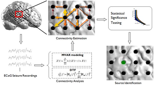
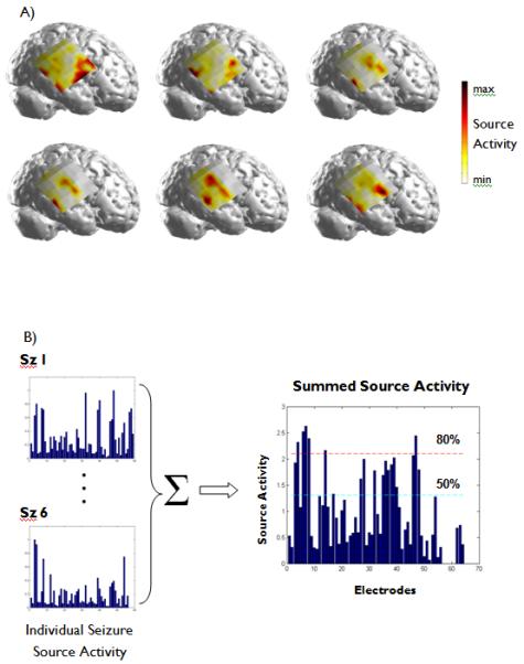
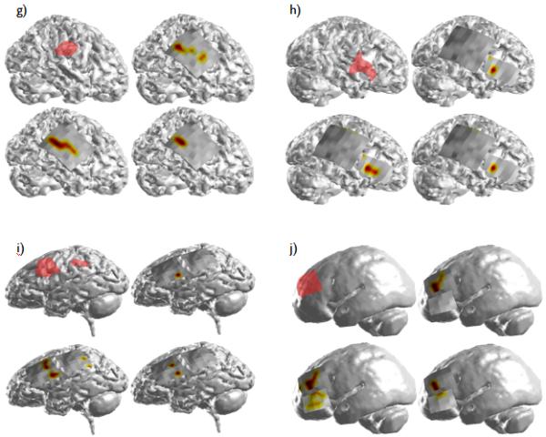
Methods: We applied a causal measurement technique--the directed transfer function (DTF)--in an effort to determine the cortical location responsible for the propagation of the seizure activity. Intracranial seizure recordings were obtained from a group of 11 pediatric patients with medically intractable neocortical-onset epilepsy. Time windows were selected from the recordings following onset of the ictal activity. The DTF was applied to the selected time windows, and the frequency-specific statistically significant source activity arising from each cortical recording site was quantified. The DTF-estimated source activity was then compared with the seizure-onset zone(s) identified by the epileptologists.
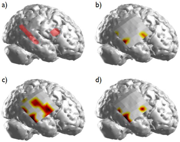
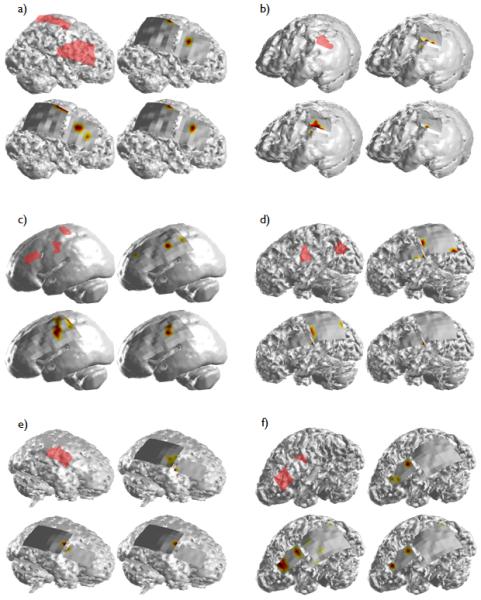
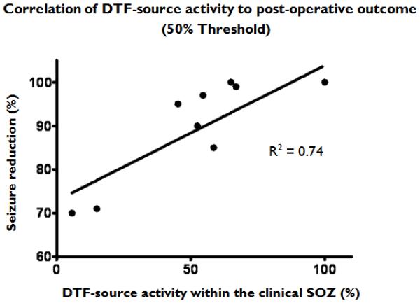
Results: In an analysis of the 11 pediatric patients, the DTF was shown to identify estimated ictal sources that were highly correlated with the clinically identified foci. In addition, it was observed that in the patients with multiple ictal foci, the topography of the casual source activity from the analyzed seizures was associated with the separate clinically identified seizure-onset zones.
Discussion: Although localization of neocortical-onset seizures is typically challenging, the causal measures employed in this study-namely the directed transfer function-identified generators of the ictal activity that were highly correlated with the cortical regions identified as the seizure-onset zones by the epileptologists. This technique could prove useful in the identification of seizure-specific propagation pathways in the presurgical evaluation of patients with epilepsy.
Figures
Similar articles
-
Duration of complex partial seizures: an intracranial EEG study.Epilepsia. 2008 Apr;49(4):677-84. doi: 10.1111/j.1528-1167.2007.01420.x. Epub 2007 Nov 19. Epilepsia. 2008. PMID: 18028402
-
Brain-responsive neurostimulation in patients with medically intractable seizures arising from eloquent and other neocortical areas.Epilepsia. 2017 Jun;58(6):1005-1014. doi: 10.1111/epi.13739. Epub 2017 Apr 7. Epilepsia. 2017. PMID: 28387951 Clinical Trial.
-
Dynamic changes of ictal high-frequency oscillations in neocortical epilepsy: using multiple band frequency analysis.Epilepsia. 2007 Feb;48(2):286-96. doi: 10.1111/j.1528-1167.2007.00923.x. Epilepsia. 2007. PMID: 17295622
-
A study of the dynamics of seizure propagation across micro domains in the vicinity of the seizure onset zone.J Neural Eng. 2015 Aug;12(4):046016. doi: 10.1088/1741-2560/12/4/046016. Epub 2015 Jun 10. J Neural Eng. 2015. PMID: 26061006 Free PMC article.
-
Hypothalamic hamartoma: Epileptogenesis beyond the lesion?Epilepsia. 2017 Jun;58 Suppl 2:32-40. doi: 10.1111/epi.13755. Epilepsia. 2017. PMID: 28591482 Review.
Cited by
-
Seizure detection using the phase-slope index and multichannel ECoG.IEEE Trans Biomed Eng. 2012 Apr;59(4):1125-34. doi: 10.1109/TBME.2012.2184796. Epub 2012 Jan 18. IEEE Trans Biomed Eng. 2012. PMID: 22271828 Free PMC article.
-
Weighted and directed interactions in evolving large-scale epileptic brain networks.Sci Rep. 2016 Oct 6;6:34824. doi: 10.1038/srep34824. Sci Rep. 2016. PMID: 27708381 Free PMC article.
-
Neuroimaging in Pediatric Epilepsy.Brain Sci. 2019 Aug 7;9(8):190. doi: 10.3390/brainsci9080190. Brain Sci. 2019. PMID: 31394851 Free PMC article.
-
Connectomics and epilepsy.Curr Opin Neurol. 2013 Apr;26(2):186-94. doi: 10.1097/WCO.0b013e32835ee5b8. Curr Opin Neurol. 2013. PMID: 23406911 Free PMC article.
-
Noninvasive Electromagnetic Source Imaging and Granger Causality Analysis: An Electrophysiological Connectome (eConnectome) Approach.IEEE Trans Biomed Eng. 2016 Dec;63(12):2474-2487. doi: 10.1109/TBME.2016.2616474. Epub 2016 Oct 11. IEEE Trans Biomed Eng. 2016. PMID: 27740473 Free PMC article.
References
-
- Allen PJ, Fish DR, Smith SJ. Very high-frequency rhythmic activity during SEEG suppression in frontal lobe epilepsy. Electroencephalogr Clin Neurophysiol. 1992;82:155–159. - PubMed
-
- Astolfi L, Cincotti F, Mattia D, Salinari S, Babiloni C, Basilisco A, Rossini PM, Ding L, Ni Y, He B, Marciani MG, Babiloni F. Estimation of the effective and functional human cortical connectivity with structural equation modeling and directed transfer function applied to high-resolution EEG. Magn Reson Imaging. 2004;22:1457–1470. - PubMed
-
- Babiloni F, Cincotti F, Babiloni C, Carducci F, Mattia D, Astolfi L, Basilisco A, Rossini PM, Ding L, Ni Y, Cheng J, Christine K, Sweeney J, He B. Estimation of the cortical functional connectivity with the multimodal integration of high-resolution EEG and fMRI data by directed transfer function. NeuroImage. 2005;24:118–131. - PubMed
-
- Baccala LA, Alvarenga MY, Sameshima K, Jorge CL, Castro LH. Graph theoretical characterization and tracking of the effective neural connectivity during episodes of mesial temporal epileptic seizure. Journal of integrative neuroscience. 2004;3:379–395. - PubMed
-
- Blumenfeld H, Westerveld M, Ostroff RB, Vanderhill SD, Freeman J, Necochea A, Uranga P, Tanhehco T, Smith A, Seibyl JP, Stokking R, Studholme C, Spencer SS, Zubal IG. Selective frontal, parietal, and temporal networks in generalized seizures. NeuroImage. 2003;19:1556–1566. - PubMed
Publication types
MeSH terms
Grants and funding
LinkOut - more resources
Full Text Sources
Other Literature Sources
