

DNA damage induced by cis- and carboplatin as indicator for in vitro sensitivity of ovarian carcinoma cells
- PMID: 19818145
- PMCID: PMC2768745
- DOI: 10.1186/1471-2407-9-359
DNA damage induced by cis- and carboplatin as indicator for in vitro sensitivity of ovarian carcinoma cells
Abstract
Background: The DNA damage by platinum cytostatics is thought to be the main cause of their cytotoxicity. Therefore the measurement of the DNA damage induced by cis- and carboplatin should reflect the sensitivity of cancer cells toward the platinum chemotherapeutics.
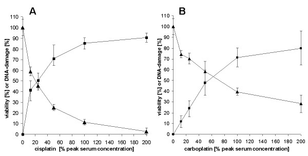
Methods: DNA damage induced by cis- and carboplatin in primary cells of ovarian carcinomas was determined by the alkaline comet assay. In parallel, the reduction of cell viability was measured by the fluorescein diacetate (FDA) hydrolysis assay.
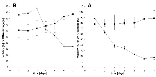
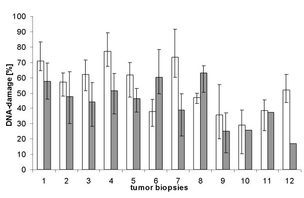
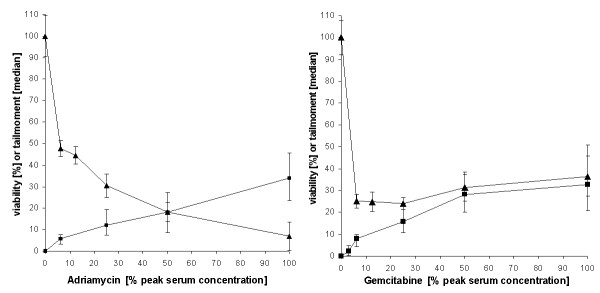
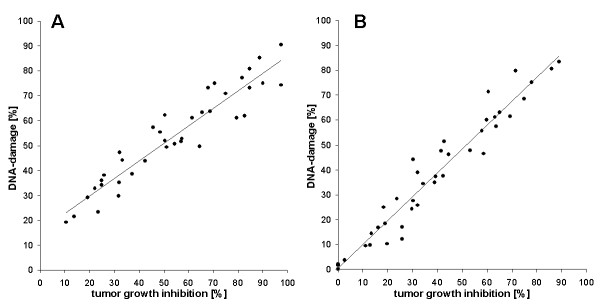
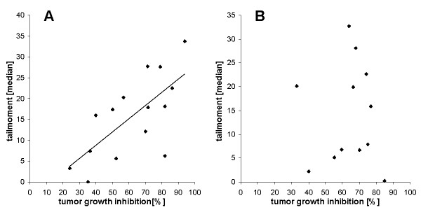
Results: While in the comet assay the isolated cells showed a high degree of DNA damage after a 24 h treatment, cell viability revealed no cytotoxicity after that incubation time. The individual sensitivities to DNA damage of 12 tumour biopsies differed up to a factor of about 3. DNA damage after a one day treatment with cis- or carboplatin correlated well with the cytotoxic effects after a 7 day treatment (r = 0,942 for cisplatin r = 0.971 for carboplatin). In contrast to the platinum compounds the correlation of DNA damage and cytotoxicity induced by adriamycin was low (r = 0,692), or did not exist for gemcitabine.
Conclusion: The measurement of DNA damage induced by cis- and carboplatin is an accurate method to determine the in vitro chemosensitivity of ovarian cancer cells towards these cytostatics, because of its quickness, sensitivity, and low cell number needed.
Figures
Similar articles
-
Antitumor carboplatin is more toxic in tumor cells when photoactivated: enhanced DNA binding.J Biol Inorg Chem. 2012 Aug;17(6):891-8. doi: 10.1007/s00775-012-0906-z. Epub 2012 May 26. J Biol Inorg Chem. 2012. PMID: 22638735
-
Chemoresistance testing of human ovarian cancer cells and its in vitro model.Toxicol In Vitro. 2010 Dec;24(8):2108-15. doi: 10.1016/j.tiv.2010.08.010. Epub 2010 Aug 22. Toxicol In Vitro. 2010. PMID: 20736059
-
Chloroxine overrides DNA damage tolerance to restore platinum sensitivity in high-grade serous ovarian cancer.Cell Death Dis. 2021 Apr 14;12(4):395. doi: 10.1038/s41419-021-03665-0. Cell Death Dis. 2021. PMID: 33854036 Free PMC article.
-
Selective inhibition of tumor cell associated Vacuolar-ATPase 'a2' isoform overcomes cisplatin resistance in ovarian cancer cells.Mol Oncol. 2016 Jun;10(6):789-805. doi: 10.1016/j.molonc.2016.01.003. Epub 2016 Jan 29. Mol Oncol. 2016. PMID: 26899534 Free PMC article.
-
Medical therapy of advanced malignant epithelial tumours of the ovary.Forum (Genova). 2000 Oct-Dec;10(4):323-32. Forum (Genova). 2000. PMID: 11535983 Review.
Cited by
-
Phase I Trial of Carboplatin and Gemcitabine Chemotherapy and Stereotactic Ablative Radiosurgery for the Palliative Treatment of Persistent or Recurrent Gynecologic Cancer.Front Oncol. 2015 Jun 5;5:126. doi: 10.3389/fonc.2015.00126. eCollection 2015. Front Oncol. 2015. PMID: 26097831 Free PMC article.
-
Circulating non‑coding RNA‑biomarker potential in neoadjuvant chemotherapy of triple negative breast cancer?Int J Oncol. 2020 Jan;56(1):47-68. doi: 10.3892/ijo.2019.4920. Epub 2019 Nov 25. Int J Oncol. 2020. PMID: 31789396 Free PMC article.
-
Enhanced sensitivity to cisplatin and gemcitabine in Brca1-deficient murine mammary epithelial cells.BMC Pharmacol. 2011 Jul 19;11:7. doi: 10.1186/1471-2210-11-7. BMC Pharmacol. 2011. PMID: 21771338 Free PMC article.
-
Human mesenchymal stem cells are resistant to cytotoxic and genotoxic effects of cisplatin in vitro.Genet Mol Biol. 2016 Mar;39(1):129-34. doi: 10.1590/1678-4685-GMB-2015-0057. Genet Mol Biol. 2016. PMID: 27007906 Free PMC article.
-
Evaluation for Synergistic Effects by Combinations of Photodynamic Therapy (PDT) with Temoporfin (mTHPC) and Pt(II) Complexes Carboplatin, Cisplatin or Oxaliplatin in a Set of Five Human Cancer Cell Lines.Int J Mol Sci. 2018 Oct 16;19(10):3183. doi: 10.3390/ijms19103183. Int J Mol Sci. 2018. PMID: 30332729 Free PMC article.
References
-
- Gonzalez VM, Fuertes MA, Alonso C, Perez JM. Is cisplatin-induced cell death always produced by apoptosis? Mol Pharmacol. 2001;59:657–663. - PubMed
Publication types
MeSH terms
Substances
LinkOut - more resources
Full Text Sources
Medical
