. 2010 Jan;117(1):133-139.e2.
doi: 10.1016/j.ophtha.2009.05.029. Epub 2009 Oct 8.

Rituximab treatment of patients with severe, corticosteroid-resistant thyroid-associated ophthalmopathy

Affiliations

Rituximab treatment of patients with severe, corticosteroid-resistant thyroid-associated ophthalmopathy

Dinesh Khanna et al. Ophthalmology. 2010 Jan.

Abstract

Purpose: To study the effectiveness of anti-CD20 (rituximab [RTX]; Rituxan; Genentech, Inc., South San Francisco, CA) therapy in patients with severe, corticosteroid (CS)-resistant thyroid-associated ophthalmopathy (TAO).

Design: Retrospective, interventional case series.

Participants: Six consecutive subjects with severe, progressive TAO unresponsive to CS.

Methods: Electronic medical record review of consecutive patients receiving RTX during the previous 18 months. Responses to therapy were graded using standard clinical assessment and flow cytometric analysis of peripheral lymphocytes.

Main outcome measures: Clinical activity score (CAS), proptosis, strabismus, treatment side effects, and quantification of regulatory T cells.

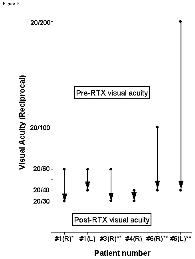
Results: Six patients were studied. Systemic CS failed to alter clinical activity in all patients (mean CAS+/-standard deviation, 5.3+/-1.0 before vs. 5.5+/-0.8 during therapy for 7.5+/-6.4 months; P = 1.0). However, after RTX treatment, CAS improved from 5.5+/-0.8 to 1.3+/-0.5 at 2 months after treatment (P<0.03) and remained quiescent in all patients (CAS, 0.7+/-0.8; P<0.0001) at a mean follow-up of 6.2+/-4.5 months. Vision improved bilaterally in all 4 patients with dysthyroid optic neuropathy (DON). None of the 6 patients experienced disease relapse after RTX infusion, and proptosis remained stable (Hertel measurement, 24+/-3.7 mm before therapy and 23.6+/-3.7 mm after therapy; P = 0.17). The abundance of T regulatory cells, assessed in 1 patient, increased within 1 week of RTX and remained elevated at 18 months of follow-up.

Conclusions: In progressive, CS-resistant TAO, rapid and sustained resolution of orbital inflammation and DON followed treatment with RTX.

Financial disclosure(s): The author(s) have no proprietary or commercial interest in any materials discussed in this article.


Figures

Figure 1
Figure 1. Clinical response after Rituximab (RTX) infusion
A) Clinical activity score (CAS) of thyroid associated ophthalmopathy (TAO) evaluated before and after infusion of RTX, wk = weeks. * denotes patients who developed dysthyroid optic neuropathy (DON) during corticosteroid (CS) treatment. B) Summary of CAS before and after CS and RTX therapy in all six patients. *p<0.05(after CS and after RTX at 2 months), Wilcoxon signed rank test **p<0.0001(after CS, after RTX at 2 months and at last follow-up), Friedman test C) Change in visual acuity (reciprocal) in subjects with DON before and after RTX infusion. Abscissa represents individual eye, top of arrow indicates initial visual acuity, arrowhead points to final visual acuity. (*)Final visual acuity assessed prior to transcaruncular medial wall orbital decompression. (**)Initial visual acuity assessed after transcaruncular medial wall orbital decompression but prior to RTX infusion. D) Assessment of proptosis (mean + standard deviation) before and after RTX. Measure of proptosis was performed before and after RTX in the unoperated orbit for patients 1–5 and the worse side for patient 6. P=0.17, Wilcoxon signed rank test
Figure 2
Figure 2
Photographs of two patients demonstrating orbital inflammation before and after Rituximab (RTX) therapy. Patient 4 demonstrated significant periorbital inflammation (A) prior to RTX treatment which decreased dramatically after therapy (B). Stabismsus and eyelid retraction increased despite improvement of inflammatory signs and symptoms. Patient 1 prior to RTX treatment (C) and approximately 18 months after treatment (D).
Figure 3
Figure 3. Histology and immunohistochemical staining of orbital tissues obtained during decompression surgery from patient 1
A) High-powered view demonstrating adipocytes with prominent fibrovascular septae and paucity of mononuclear inflammatory infiltrates. B) High-powered view demonstrating absence of CD20+ B cells and C) High-powered view demonstrating absence of CD3+ T cells A) Hematoxylin and Eosin. B & C) counterstained with Hematoxylin. Magnification, x400, (inset, x40).
Figure 4
Figure 4
Flow cytometric analysis of regulatory T (Treg) cells in Patient 1 before the initiation of therapy, 2 weeks and 18 months after Rituximab (RTX) infusion. The data show increased Treg (CD25Hi) cells among CD4+ T cells.

Comment in

  • Thyroid-associated orbitopathy.
    Tarabishy AB, Lewis CD, Perry JD. Tarabishy AB, et al. Ophthalmology. 2010 Sep;117(9):1864; author reply 1864-5. doi: 10.1016/j.ophtha.2010.05.006. Ophthalmology. 2010. PMID: 20816259 No abstract available.
  • Rituximab for thyroid eye disease.
    Oh SR, Priel A, Levi L, Granet DB, Korn BS, Kikkawa DO. Oh SR, et al. Ophthalmology. 2011 Apr;118(4):792; author reply 792-3. doi: 10.1016/j.ophtha.2011.01.012. Ophthalmology. 2011. PMID: 21459230 No abstract available.

References

    1. Jacobson DL, Gange SJ, Rose NR, Graham NM. Epidemiology and estimated population burden of selected autoimmune diseases in the United States. Clin Immunol Immunopathol. 1997;84:223–243. - PubMed
    1. McGrogan A, Seaman HE, Wright JW, de Vries CS. The incidence of autoimmune thyroid disease: a systematic review of the literature. Clin Endocrin (Oxf) 2008;69:687–696. - PubMed
    1. Weetman AP. Graves' disease. N Engl J Med. 2000;343:1236–1248. - PubMed
    1. Smith TJ, Tsai CC, Shih MJ, et al. Unique attributes of orbital fibroblasts and global alterations in IGF-1 receptor signaling could explain thyroid-associated ophthalmopathy. Thyroid. 2008;18:983–988. - PMC - PubMed
    1. Douglas RS, Gianoukakis AG, Goldberg RA, et al. Circulating mononuclear cells from euthyroid patients with thyroid-associated ophthalmopathy exhibit characteristic phenotypes. Clin Exp Immunol. 2007;148:64–71. - PMC - PubMed

Publication types

MeSH terms