Review
. 2009 Dec;34(12):628-39.
doi: 10.1016/j.tibs.2009.08.002. Epub 2009 Oct 7.

Lafora disease: insights into neurodegeneration from plant metabolism

Affiliations
Review

Lafora disease: insights into neurodegeneration from plant metabolism

Matthew S Gentry et al. Trends Biochem Sci. 2009 Dec.

Abstract

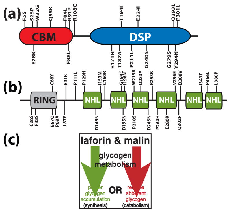
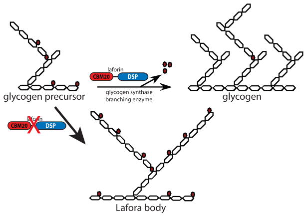
Reversible phosphorylation modulates nearly every step of glycogenesis and glycogenolysis. Multiple metabolic disorders are the result of defective enzymes that control these phosphorylation events, enzymes that were identified biochemically before the advent of the molecular biology era. Lafora disease is a metabolic disorder resulting in accumulation of water-insoluble glucan in the cytoplasm, and manifests as a debilitating neurodegeneration that ends with the death of the patient. Unlike most metabolic disorders, the link between Lafora disease and metabolism has not been defined in almost 100 years. The results of recent studies with mammalian cells, mouse models, eukaryotic algae, and plants have begun to define the molecular mechanisms that cause Lafora disease. The emerging theme identifies a new phosphorylation substrate in glycogen metabolism, the glucan itself.


Figures

Figure 1
Figure 1
A schematic of laforin and malin. Amino acid substitutions stemming from Lafora disease missense mutations are shown for laforin and malin. (a) Laforin contains a carbohydrate binding module (CBM) and a dual specificity phosphatase domain (DSP). (b) Malin contains a RING domain followed by six NHL repeats. (c) Malin and laforin are involved in one of the two branches of glycogen metabolism.
Figure 2
Figure 2
Models of glycogen and amylopectin. A three dimensional structure of glycogen and starch cannot be determined experimentally due to their polydispersity, but these are the widely accepted models [–100]. In each model, solid lines represent glucan chains. (a) Glycogen production is initiated when glycogenin (G) covalently attaches glucose to itself at Tyr194 and continues with the autocatalytic addition of about 10 glucosyl residues. This protein–glucosyl complex serves as the starting point that glycogen synthase and branching enzyme utilize to link glucose by α-1,4-glycosidic linkages with branches linked by α-1,6-glycosidic linkages every 12–14 residues. Glycogen synthase and branching enzyme construct up to twelve tiers of branches, five of which are depicted here. These tiers are organized in a continuous manner, rendering glycogen water soluble. (b) Amylopectin is also composed of α-1,4-glycosidic linkages with α-1,6-glycosidic branches, but with branches arranged in clusters at regular intervals. The glucan chains within the clusters interact and this is represented by intersection of the adjacent chains, which form double helices and organize into crystalline lamellae. Between each cluster is a non-branched region that makes up the amorphous lamellae. The decreased branching and the crystalline lamellae render amylopectin, and starch, water insoluble.
Figure 3
Figure 3
Model of Lafora body formation caused by loss of laforin. Glucose moieties are depicted as hexagons. Glucose is linked by α-1,4-glycosidic linkages with branches via α-1,6-glycosidic linkages. Glycogen contains small amounts of covalently linked phosphate (0.25% w/w), present as both phosphomonoesters and phosphodiesters [–43, 65]. Phosphate is represented by red-filled circles, with phosphomonesters adjacent glucose hexagons and phosphodiesters between two glucose hexagons. As nascent glycogen molecules are being synthesized by glycogen synthase and branching enzyme, phosphomonoesters and phosphodiesters accumulate by an unknown mechanism. Laforin removes phosphomonoesters so that glycogen production proceeds normally. In the absence of laforin, phosphomonoesters accumulate and negatively impact glycogen branching and lead to Lafora body (LB) formation. LBs contain increased amounts of phosphate and decreased branching compared to glycogen, and these two characteristics make LBs insoluble.
Figure 4
Figure 4
Structural and bioinformatic properties of laforin. (a) Slices of the active site surface of three classes of PTPs: 1) the deep and wide active site of the phosphoinositol phosphatase MTMR2 in blue, 2) the deep and narrow active site of the pTyr-specific phosphatase PTP1B in green, and 3) the shallow and narrow active site of the dual-specific phosphatase VHR in orange [101]. Used with permission from Current Opinion in Structural Biology. (b) An alignment and secondary structure prediction of human laforin and VHR (hVHR) were generated using PROMALS [102]. The accepted phosphatase motifs are indicated above each segment and the secondary structure is indicated below. Similar amino acids are boxed in light grey and identical amino acids in dark grey. The recognition domain and variable loop are highlighted in green and red, respectively. (c) A phylogeny built using the catalytic domain of the dual specificity phosphatases. The more recently evolved MAPK phosphatases or “classical” DSPs are highlighted with a grey background. The more ancient and divergent “atypical” DSPs fall into two groups. One group is relatively tightly clustered and utilizes proteinaceous substrates and this group has no highlighted background. The second group is more divergent and has a tan background, and includes laforin and SEX4. Most of the DSPs within this clade dephosphorylate non-proteinaceous substrates (e.g. phosphoinositols, RNA, and glucans), highlighted in green boxes. Some of the DSPs in ths clade have undefined in vivo substrates, but they have activity against non-proteinaceous substrates in vitro, highlighted with a green dashed line. The phylogenetic tree was generated from a PROMALS multiple sequence alignment using PROTDIST and FITCH from the PHYLIP 3.65 software package and displayed using HYPERTREE 1.0.0 [102, 103].
Figure 5
Figure 5
Evolutionary conservation of laforin and SEX4. (a) Schematic of SEX4. SEX4 is composed of a chloroplast targeting peptide (cTP), dual specificity phosphatase domain (DSP), and carbohydrate binding module family 21 (CBM21). (b) Unrooted phylogeny of the small subunit ribosomal RNA (SSU rRNA) from organisms representing many evolutionary niches (modified [78]). Organisms containing laforin are boxed in yellow and those containing SEX4 are boxed in green. Alveolates are shaded with a grey background and vertebrates with a brown background. Bootstrap values are indicated by color coding in the inset. (c) Unrooted phylogeny of all SEX4 orthologs. Bootstrap values are as in b. The phylogenetic trees were generated as in Figure 4. Double hash marks indicate a place where intervening segment was removed due to space limitations.
Figure 6
Figure 6
Models depicting the role of laforin and SEX4 in glucan metabolism. (a) Proposed model of starch breakdown (Copyright American Society of Plant Biologists, www.plantcell.org) [64]. Starch is phosphorylated (red circles) at night by GWD and PWD (i), leading to unwinding of amylopectin double helices. In WT plants, β-amylase isozyme 3 (BAM3) and SEX4 release maltose and phosphate, respectively (ii), and isoamylase 3 (ISA3) hydrolyzes branch points and releases malto-oligosaccharides (iii). In sex4 mutants, phosphate is not hydrolyzed by SEX4, leading to reduced maltose release by BAM3 (iv). Subsequently, α-amylase (AMY3) and ISA3 release both malto- and phospho-oligosaccharides. Following degradation of the outer layer, a new round of degradationcan begin with the phosphorylation of the granule surface by GWD and PWD (i). (b) We propose that in plants and protists, laforin and SEX4 are involved in converting insoluble glucans into usable energy. (c) In humans, all other vertebrates, and at least two invertebrates (Nematostella and Branchiostoma), laforin inhibits insoluble glucan accumulation by dephosphorylating nascent glycogen molecules as proposed in Figure 3. The photograph of Dr. Gonzalo Rodriguez Lafora was and the image of a Tetrahymena was reproduced with permission [104]. All other images were generated by the authors or obtained from non-restricted copyright sources.

References

    1. Lafora G, Glick G. Beitrag zur histopathologie der myoklonischen epilepsie. Z Ges Neurol Psychiatr. 1911;6:1–14.
    1. Lafora GR. Uber des Vorkommen amyloider KJrperchen im innern der Ganglienzellen. Virchows Arch f Path Anat. 1911;205:295.
    1. Virchow RLK. Die Cellularpathologie inihrer Begründung auf physiologische and pathologische Gewebelehre. Berlin: Hirschwald; 1858.
    1. Yokoi S, Austin J, Witmer F. Isolation and characterization of Lafora bodies in two cases of myoclonus epilepsy. Journal of Neuropathology and Experimental Neurology. 1967;26(1):125–127. - PubMed
    1. Yokoi S, Austin J, Witmer F, Sakai M. Studies in myoclonus epilepsy (Lafora body form). I. Isolation and preliminary characterization of Lafora bodies in two cases. Arch Neurol. 1968;19(1):15–33. - PubMed

Publication types

MeSH terms

Substances