. 2009 Oct 9;36(1):39-50.
doi: 10.1016/j.molcel.2009.09.022.

Structures of SPOP-substrate complexes: insights into molecular architectures of BTB-Cul3 ubiquitin ligases

Affiliations

Structures of SPOP-substrate complexes: insights into molecular architectures of BTB-Cul3 ubiquitin ligases

Min Zhuang et al. Mol Cell. .

Abstract

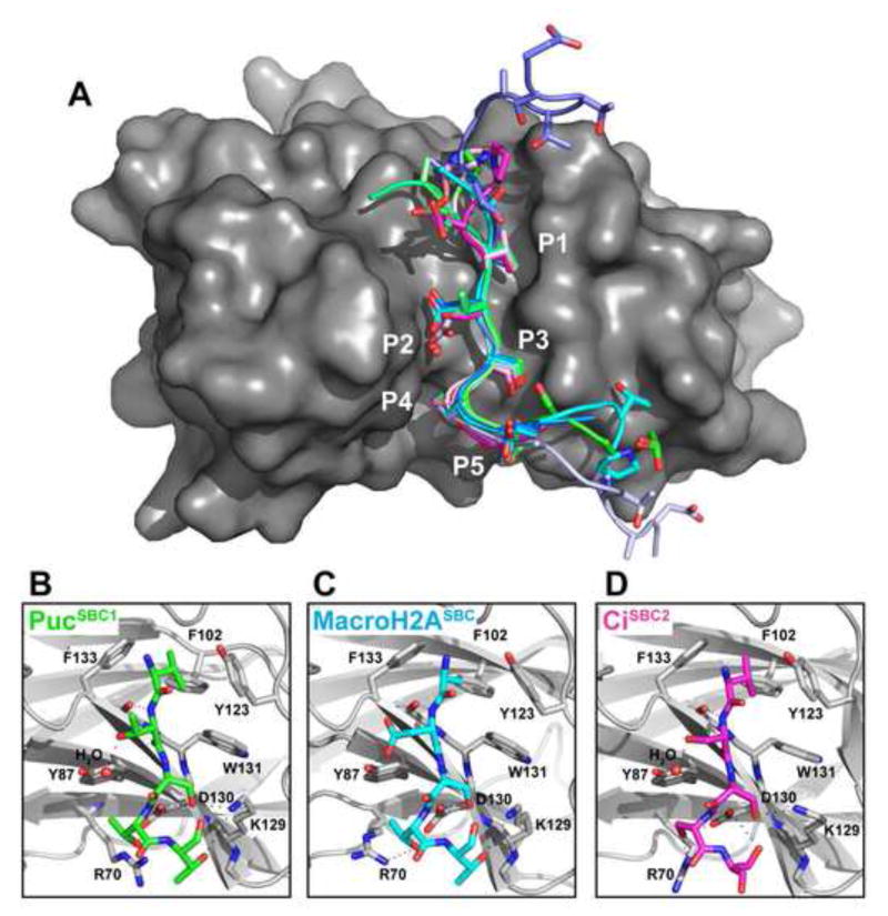
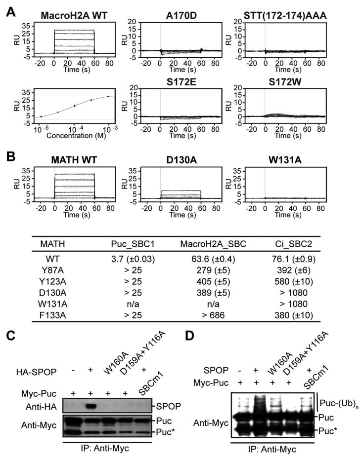
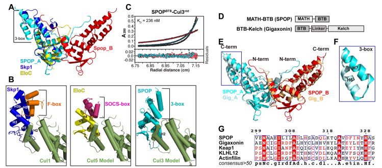
In the largest E3 ligase subfamily, Cul3 binds a BTB domain, and an associated protein-interaction domain such as MATH recruits substrates for ubiquitination. Here, we present biochemical and structural analyses of the MATH-BTB protein, SPOP. We define a SPOP-binding consensus (SBC) and determine structures revealing recognition of SBCs from the phosphatase Puc, the transcriptional regulator Ci, and the chromatin component MacroH2A. We identify a dimeric SPOP-Cul3 assembly involving a conserved helical structure C-terminal of BTB domains, which we call "3-box" due to its facilitating Cul3 binding and its resemblance to F-/SOCS-boxes in other cullin-based E3s. Structural flexibility between the substrate-binding MATH and Cul3-binding BTB/3-box domains potentially allows a SPOP dimer to engage multiple SBCs found within a single substrate, such as Puc. These studies provide a molecular understanding of how MATH-BTB proteins recruit substrates to Cul3 and how their dimerization and conformational variability may facilitate avid interactions with diverse substrates.


Figures

Figure 1
Figure 1. Identification of a SPOP Binding Consensus (SBC) sequence in multiple SPOP substrates
(A) Identification of a SPOP-binding peptide in Puc. Left, Coommassie-stained SDS-PAGE gel showing products after trypsin digestion of a complex between a SPOP MATH domain and Puc1-390 (333:1 trypsin, 3 hr, rt). Right, Coommassie-stained SDS-PAGE gel of fractions from gel filtration (SD200) of trypsin digest products. Bottom, peptide co-purifying with SPOPMATH in fractions 33–35, identified by mass spectrometry. (B) Identification of a MacroH2A sequence required for binding to SPOP. Top, Schematic view of MacroH2A deletions, highlighting residues 166-179. Bottom, Coommassie-stained SDS-PAGE gel of GST-pull-downs of GST, GST-MacroH2A 1, and GST-MacroH2A 2 coexpressed in E. coli with a HisMBP-tagged SPOP MATH domain. (C) SBCs (red) in SPOP substrates Puc, MacroH2A, Ci and Daxx. (D) Binding constants for SPOPMATH interactions with SBC peptides, measured by Surface Plasmon Resonance (BIACORE3000). (E) Roles of individual Puc SBCs in in vitro ubiquitination. Western blots detecting His-Puc ubiquitination for wild-type (WT) and mutant Puc substrates. SBCm1, SBCm2 and SBCm3 refer to mutation at the 3 SBC sites.
Figure 2
Figure 2. Structural Basis for SPOPMATH-SBC Interactions
(A) Comparison of 11 independent structures of isolated SPOPMATH complexed with SBC peptides (3 from Puc – greens; 4 from MacroH2A – cyans/blues; 4 from Ci – pinks/magentas). After superposition over SPOPMATH mainchain, SBC peptides were displayed with backbones as cartoons and sidechains as sticks, docked in the structure of SPOPMATH (grey surface) from the complex with PucSBC1. The 5 φ-π-S-S/T-S/T motif positions are indicated P1–P5. (B–D) Close-up views of SPOPMATH (grey) complexes with (B) PucSBC1 (green), (C) MacroH2ASBC (cyan), and (D) CiSBC2 (magenta), oriented as in panel A. Dashed lines - hydrogen bonds; red – oxygen; blue – nitrogen; red sphere – water.
Figure 3
Figure 3. Mutational Analysis of SPOPMATH-Substrate Interaction
(A) BIACORE sensograms showing SPOPMATH binding to wildtype or indicated mutant versions of a MacroH2ASBC peptide. Bottom left - Fit used to calculate KD for SPOPMATH-MacroH2ASBC. (B) Representative BIACORE sensograms examining binding between wildtype or indicated mutant versions of SPOPMATH and a PucSBC1 peptide. BIACORE binding data for SPOPMATH mutants and SBC peptides is summarized below. (C) Western blots showing association of HA-SPOP (top) with Myc-Puc (bottom) or mutants after anti-Myc immunoprecipitation from Drosophila S2 cells co-transfected with the indicated constructs. D130 of human SPOP corresponds to D159 in Drosophila SPOP, W131 corresponds to W160, and Y87 corresponds to Y116. *Processed form of Puc (Liu et al., 2009). (D) Anti-Myc western blot detecting ubiquitination of Puc from S2 cells co-transfected with wild-type or mutant versions of SPOP.
Figure 4
Figure 4. SPOPBTB+ forms a 2:2 dimer with Cul3 N-terminal domain
(A) Left, overall view of the SPOPBTB+ dimer, with protomers in cyan (A) and red (B). Right, close-up view of dimer interface rotated 90° in x. (B) Equilibrium AUC of SPOPBTB++Cul3ntd. Samples at 1.0 to 8.8 μM centrifuged at 8 (red), 12 (blue), and 16 (black) krpm 4 C. Lines show global nonlinear least-squares best-fit of all datasets/concentrations/speeds to a heterogeneous association model describing a 2:2 SPOPBTB+:Cul3ntd complex (MW 127.1 kDa) with indicated KD value. For clarity, only the 3 μM sample is shown. (C) AUC of L186D, L190D, L193D, I217K mutant SPOPBTB++Cul3ntd performed as in B. Lines show global nonlinear least-squares best-fit of all datasets/concentrations/speeds to a heterogeneous association model describing a 1:1 SPOPBTB+ (mutant):Cul3ntd complex (MW 63.6 kDa) with the indicated KD value. For clarity, only the 2.0 μM sample is shown. (D) Western blots of SPOPMATH-BTB+-mediated ubiquitination detecting His-Puc, for wild-type and L186D, L190D, L193D, I217K (dimer-defective) mutant SPOP.
Figure 5
Figure 5. A conserved Cul3-Interacting box (3-box)
(A) Structural alignment of SPOPBTB+, Skp1 (blue) and EloC (yellow). One molecule of the SPOPBTB+ dimer is cyan (SPOP_A) and one is red (SPOP_B). The SPOP helix-pair (above “+”) not shared in Skp1 and EloC is labeled “3-box”. (B) Structural comparison of the Skp1 (blue) -F-boxSkp2 (orange) -Cul1 (green) structure (1LDK.pdb), the Elongin C (yellow) – SOCS-boxVHL (magenta) structure (1VCB.pdb) docked onto a structural model of Cul5 (green), and the SPOPBTB/3-box (cyan) docked on a structural model of Cul3 (green). The relative locations of the SPOP 3-box, F-box, and SOCS-box are indicated. (C) Equilibrium AUC of SPOPBTB (i.e., lacking the 3-box)+Cul3ntd. Samples at 1.2 to 8.0 μM centrifuged at 8 (red), 12 (blue), and 15 (black) krpm 4 C. Lines represent the global nonlinear least-squares best-fit of all datasets/concentrations/speeds to a heterogeneous association model describing a 2:2 SPOPBTB:Cul3ntd complex (MW 118.6 kDa) with the indicated KD value. For clarity, only the 3.0 μM sample is shown. (D) Schematic of SPOP and Gigaxonin domain arrangements, which represent two distinct BTB subfamilies, MATH-BTB and BTB-Kelch, respectively. (E) Overall structural alignment of SPOPBTB/3-box and GigaxoninBTB/3-box. Residues C-terminal of the Gigaxonin 3-box were omitted for clarity. (F) Superposition of the SPOP and Gigaxonin 3-boxes. SPOP3-box –cyan; Gigaxonin3-box - light blue. (G) Structure based sequence alignment (ESPript) of 32 amino acids corresponding to 3-boxes from 5 Cul3-interacting BTB proteins. SPOP 3-box residue numbers are shown on top.
Figure 6
Figure 6. Crystal Structures of dimeric SPOPMATHx-BTB/3-box-PucSBC1
(A) Overall architecture of SPOPMATHx-BTB/3-box dimer. One molecule (A) is colored cyan and the other (B) is red. Each MATH domain binds one PucSBC1 (green) peptide. Disordered regions not visible in electron density are represented with dotted lines to show connectivity. (B) Superposition over a BTB domain for SPOPMATHx-BTB/3-box structures determined from crystals with slightly different unit cells. MATH_A1 (cyan) and MATH_B1 (red) are from crystal form 1, and correspond to the structure in (A). MATH_A2 (orange) and MATH_B2 (blue) are from crystal form 2. (C) Coommassie-stained SDS-PAGE gel showing products of proteolysis of SPOPMATH-BTB/3-box by Endoproteinase Glu-C at room temperature for 30 min. The identities of products determined by Mass Spectrometry are shown. (D) SPOPMATH and SPOPBTB/3-box domains do not cofractionate by sizing. A thrombin cleavage site was engineered in the interdomain linker in SPOPMATH-BTB/3-box (−T), and after treatment with thrombin (+T), the product was subject to gel filtration chromatography. Individual fractions were analyzed by Coommassie-stained SDS-PAGE gel, below.
Figure 7
Figure 7. A 1:2 substrate complex with the SPOP-Cul3 ubiquitin ligase
(A) Velocity AUC of SPOPMATH-BTB/3-box + Puc1-390 at 20 °C, 60 krpm fit to a continuous distribution model c(s). Two peaks indicate molecular weights of 110 kDa and 39 kDa corresponding to the 1:2 Puc:SPOPMATH-BTB/3-box complex (MWcalc 112.5 kDa) and excess free Puc (MWcalc of 42.1 kDa). (B) Equilibrium AUC of a sample as in (A). Samples at 1 to 6 μM centrifuged at 8 (red), 12 (blue), and 16 (black) krpm 4 C. Lines show global nonlinear least squares best-fit of all datasets/concentrations/speeds to a heterogeneous association model with 2 species, 2:1 SPOPMATH-BTB/3-box:Puc + Puc. For clarity, only the 1.1 μM sample is shown. (C) Overall structure of SPOPMATHx-MacroH2ASBC (pep2). Two isolated MATH domains (chain A, cyan; chain B, pink) bind a single substrate peptide (green) at two suboptimal SBC sites. (D) Schematic view of a SPOP-Cul3 ubiquitin ligase bound to a single substrate. Substrate is shown in grey, with SBCs in green, and ubiquitin-acceptor lysines as Ks. The two protomers of the dimeric SPOP complex are shown in cyan and red, with each BTB/3-box bound near the N-terminus of an elongated Cul3 (olive) activated with NEDD8 (orange) near the C-terminus. E2-bound Rbx1 RING domains are shown flexibly tethered to the Cul3 C-terminal domains. The high degree of conformational flexibility may allow substrates with a range of SBC configurations to be polyubiquitinated at multiple sites.

References

    1. Allen E, Ding J, Wang W, Pramanik S, Chou J, Yau V, Yang Y. Gigaxonin-controlled degradation of MAP1B light chain is critical to neuronal survival. Nature. 2005;438:224–228. - PubMed
    1. Angers S, Li T, Yi X, MacCoss MJ, Moon RT, Zheng N. Molecular architecture and assembly of the DDB1-CUL4A ubiquitin ligase machinery. Nature. 2006a;443:590–593. - PubMed
    1. Angers S, Thorpe CJ, Biechele TL, Goldenberg SJ, Zheng N, MacCoss MJ, Moon RT. The KLHL12-Cullin-3 ubiquitin ligase negatively regulates the Wnt-beta-catenin pathway by targeting Dishevelled for degradation. Nat Cell Biol. 2006b;8:348–357. - PubMed
    1. Bai C, Sen P, Hofmann K, Ma L, Goebl M, Harper JW, Elledge SJ. SKP1 connects cell cycle regulators to the ubiquitin proteolysis machinery through a novel motif, the F-box. Cell. 1996;86:263–274. - PubMed
    1. Bardwell VJ, Treisman R. The POZ domain: a conserved protein-protein interaction motif. Genes Dev. 1994;8:1664–1677. - PubMed

Publication types

MeSH terms

Substances