. 2009 Oct 9;36(1):51-60.
doi: 10.1016/j.molcel.2009.09.025.

Alpha4 is an essential regulator of PP2A phosphatase activity

Affiliations

Alpha4 is an essential regulator of PP2A phosphatase activity

Mei Kong et al. Mol Cell. .

Abstract

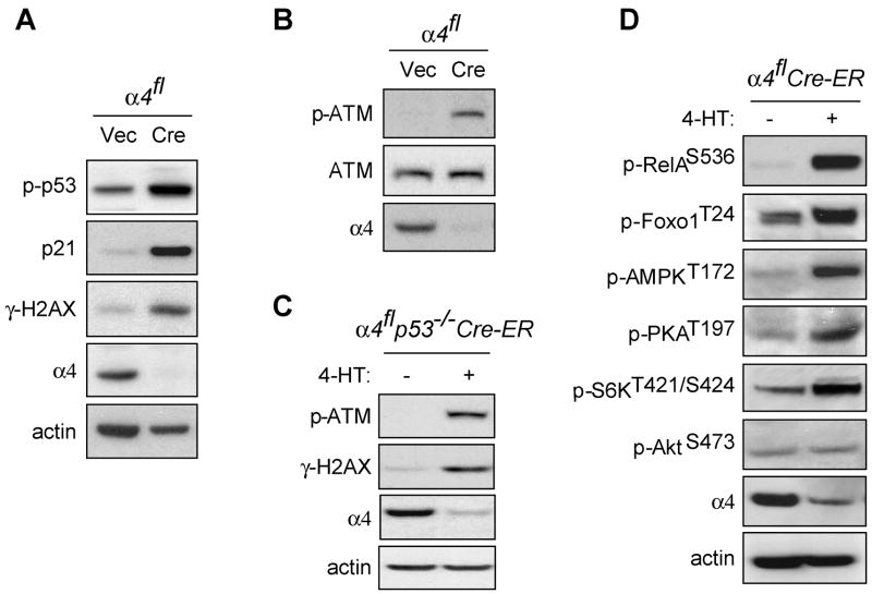
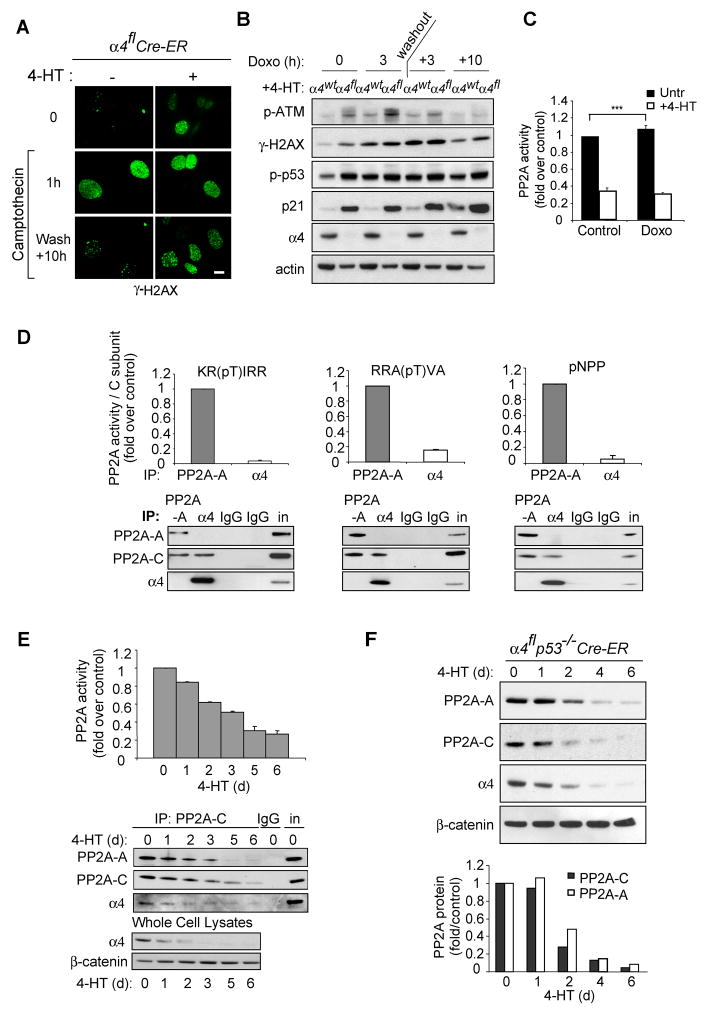
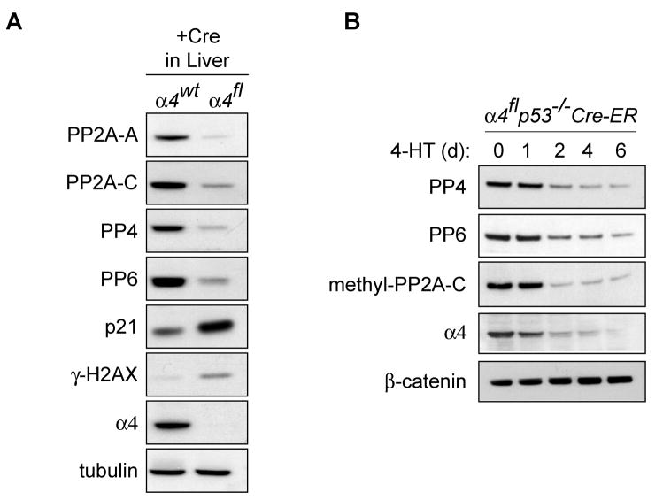
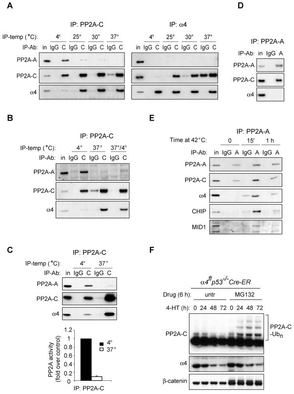
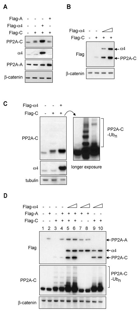
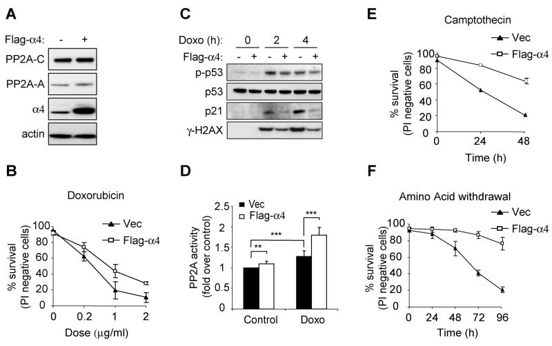
The activity and specificity of serine/threonine phosphatases are governed largely by their associated proteins. alpha4 is an evolutionarily conserved noncatalytic subunit for PP2A-like phosphatases. Though alpha4 binds to only a minority of PP2A-related catalytic subunits, alpha4 deletion leads to progressive loss of all PP2A, PP4, and PP6 phosphatase complexes. In healthy cells, association with alpha4 renders catalytic (C) subunits enzymatically inactive while protecting them from proteasomal degradation until they are assembled into a functional phosphatase complex. During cellular stress, existing PP2A complexes can become unstable. Under such conditions, alpha4 sequesters released C subunits and is required for the adaptive increase in targeted PP2A activity that can dephosphorylate stress-induced phosphorylated substrates. Consistent with this, overexpression of alpha4 protects cells from a variety of stress stimuli, including DNA damage and nutrient limitation. These findings demonstrate that alpha4 plays a required role in regulating the assembly and maintenance of adaptive PP2A phosphatase complexes.


Figures

Figure 1
Figure 1. Deletion of α4 leads to increased phosphorylation of multiple signaling proteins
A, B,α4fl MEFs were infected with MIGR1-GFP (Vec) or MIGR1-GFP-Cre (Cre) retrovirus. GFP-positive cells were sorted by flow cytometry after 24 h of infection and collected at 72 h. Immunoblotting was performed with antibodies as indicated in Experimental Procedures. The data presented are representative of at least four independent experiments. C, Tamoxifen inducible α4flp53−/− MEFs expressing Cre-ER fusion protein (α4flp53−/−Cre-ER) were treated with or without tamoxifen (4-HT) as described in Experimental Procedures. Immunoblotting was performed with antibodies as indicated. D, α4flCre-ER MEFs were treated with or without 4-HT for 48 h. Cell lysates were prepared and immunoblots probed with the indicated antibodies.
Figure 2
Figure 2. α4 is essential to maintain PP2A activity and protein levels
A, α4flCre-ER MEFs were treated with or without 4-HT and then exposed to camptothecin (2 μM) for 1 h, and then allowed to recover in fresh medium. Immunofluorescence was performed with an antibody specific for γ-H2AX at the indicated timepoints. Scale bar = 10 μm. B, α4wtCre-ER MEFs (α4wt) or α4flCre-ER MEFs (α4fl) were treated with tamoxifen (4-HT) for 48 h and exposed to doxorubicin (Doxo) (2 μg/mL) for 3 h, and then allowed to recover for the indicated timepoints. Immunoblotting was performed with antibodies as indicated. The data are representative of three independent experiments. C, α4flp53&−/−Cre-ER MEFs were treated with or without 4-HT followed by doxorubicin (2 μg/mL) for 2 h. Phosphatase activity was determined as described in Experimental Procedures, and the data presented are mean ± S.D. of three independent experiments performed in duplicate (*** P<0.005 by Student’s t-test). D, 3T3 MEFs were lysed and immunoprecipitated (IP) with the indicated antibodies. Phosphatase activity was determined using the indicated substrates, and activity was normalized for the amount of C subunit as indicated in the immunoblots. Values are means ± S.D. E, α4flp53−/−Cre-ER MEFs were treated with 4-HT for the indicated periods of time, and phosphatase activity was assayed as described in Experimental Procedures. Immunoblots show the PP2A complex immunoprecipitated by the PP2A-C antibody or total protein levels in whole cell lysates. Values are means ± S.D. F, α4flp53−/−Cre-ER MEFs were treated with 4-HT for the indicated periods of time, and lysates were analyzed by immunoblotting. The bar graph represents quantitation of immunoblots using Image J software (NIH). The data are representative of at least three independent experiments.
Figure 3
Figure 3. α4 deletion leads to loss of PP4, PP6 and methylated PP2A-C
A, Liver samples from α4wt or α4fl mice at 6 days after injection with Adeno-Cre were analyzed by immunoblotting with antibodies as indicated. B, α4flp53−/−Cre-ER MEFs were treated with 4-HT for the indicated periods of time, and lysates were analyzed by immunoblotting.
Figure 4
Figure 4. α4 can sequester and inhibit PP2A-C released from existing holoenzyme complexes
A, 3T3 MEFs were lysed and immunoprecipitated with antibodies against the PP2A C subunit, or α4, or control IgG at the indicated temperatures. In each panel, data are representative of experiments that have been replicated at least three times. B, 3T3 MEFs were lysed at 4°C and then immunoprecipitated at 4°C or 37°C. Alternatively cell lysates were incubated at 37°C for 3 h followed by immunoprecipitation at 4°C (37°/4°). C, 3T3 MEFs were lysed and immunoprecipitated with the C subunit antibody at either 4°C or 37°C. Immunoprecipitates were then used for the phosphatase assay as described in Experimental Procedures. Values are means ± S.D. D, 3T3 MEFs were lysed and immunoprecipitated with antibodies against the A subunit or control IgG at 4°C. E, 3T3 MEFs were subjected to heat shock (42°C) for the indicated amounts of time, then lysed and immunoprecipitated with antibody against the A subunit or IgG at 4°C. F, α4flp53−/−Cre-ER MEFs were treated with tamoxifen (4-HT) for the indicated periods of time, with or without proteasome inhibitor MG132 (10 μM) for the last 6 h before lysis. A–F, Immunoblots were probed with the specified antibodies and the bracket in F indicates polyubiquitination of the C subunit (PP2A-C-Ubn).
Figure 5
Figure 5. α4 promotes enhanced PP2A-C expression by preventing PP2A-C ubiquitination
A, Cells (293T) were transiently transfected with constructs containing vector, Flag-PP2A-A, Flag-PP2A-C, or Flag-α4 (2.5 μg DNA each for a total of 5 μg DNA per sample) and protein was analyzed by Western blotting at 72 h post-transfection. B, Stabilization of Flag-PP2A-C is proportional to α4 expression levels. Cells were transfected with a total of 5 μg DNA containing vector alone, Flag-PP2A-C (2.5 μg), and/or increasing amounts of Flag-α4 (1.25 – 2.5 μg), and protein was analyzed at 72 h post-transfection. C, Cells expressing Flag-PP2A-C or equal amounts of Flag-PP2A-C and Flag-α4 as in panel A were harvested at 72 h after transfection. A shorter exposure is provided to assess relative levels of PP2A-C (left upper panel) and longer exposure (right panel) reveals ubiquitinated PP2A-C levels. D, Cells were transfected with 2.5 μg of constructs encoding Flag-PP2A-C, Flag-PP2A-A, and either 1.25 or 2.5 μg of Flag-α4 as indicated together with vector control for a total of 7.5 μg DNA. The levels of all three tagged proteins were determined through recognition of the Flag epitope by Western blotting (upper panel).
Figure 6
Figure 6. α4 expression protects cells from various stress stimuli
A, 3T3 MEFs stably expressing vector or Flag-α4 were lysed and immunoblotting was performed using antibodies as indicated. In each panel, representative experiments are presented and are illustrative of at least three independent experiments. B, 3T3 MEFs expressing vector or Flag-α4 were treated with doxorubicin and viability was assessed by propidium iodide exclusion at 24 h. The data are presented as mean ± S.D. of triplicate samples in two independent experiments. C, 3T3 MEFs expressing vector (−) or Flag-α4 (+) were treated with doxorubicin (2 μg/mL). Cell lysates were prepared at 0, 2, or 4 h following treatment and immunoblots were probed with the indicated antibodies. D, Phosphatase activity was determined from immunoprecipitates of the C subunit in 3T3 MEFs expressing vector or Flag-α4 following 2 h treatment in the presence or absence of doxorubicin (Doxo, 2 μg/mL), as described in Experimental Procedures. The data presented are mean ± S.D. of three independent experiments. E–F, 3T3 MEFs expressing vector or Flag-α4 were treated with camptothecin (10 μM) or subjected to glutamine deprivation and viability was assessed by propidium iodide exclusion at the indicated timepoints. Data are representative of at least 2 independent clones for each cell type and are shown as the average of 3 experiments ± S.D. (**P<0.01, ***P<0.005 by Student’s t test).

References

    1. Arroyo JD, Hahn WC. Involvement of PP2A in viral and cellular transformation. Oncogene. 2005;24:7746–7755. - PubMed
    1. Baharians Z, Schonthal AH. Autoregulation of protein phosphatase type 2A expression. J Biol Chem. 1998;273:19019–19024. - PubMed
    1. Burma S, Chen BP, Murphy M, Kurimasa A, Chen DJ. ATM phosphorylates histone H2AX in response to DNA double-strand breaks. J Biol Chem. 2001;276:42462–42467. - PubMed
    1. Chen J, Peterson RT, Schreiber SL. Alpha 4 associates with protein phosphatases 2A, 4, and 6. Biochem Biophys Res Commun. 1998;247:827–832. - PubMed
    1. Chowdhury D, Keogh MC, Ishii H, Peterson CL, Buratowski S, Lieberman J. gamma-H2AX dephosphorylation by protein phosphatase 2A facilitates DNA double-strand break repair. Mol Cell. 2005;20:801–809. - PubMed

Publication types

MeSH terms

Substances