Review
. 2009 Nov;19(11):638-48.
doi: 10.1016/j.tcb.2009.08.009. Epub 2009 Oct 8.

Shining new light on 3D cell motility and the metastatic process

Affiliations
Review

Shining new light on 3D cell motility and the metastatic process

Paolo P Provenzano et al. Trends Cell Biol. 2009 Nov.

Abstract

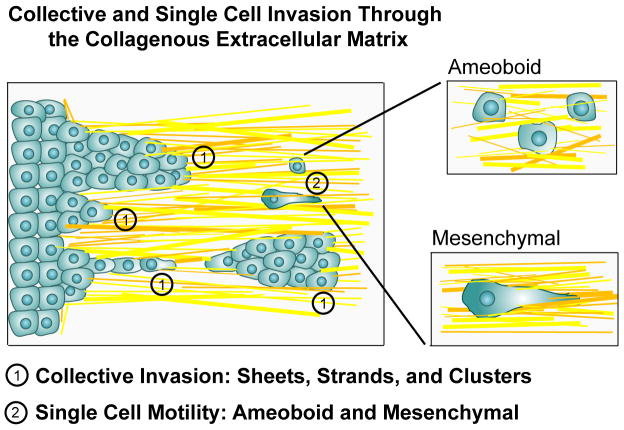
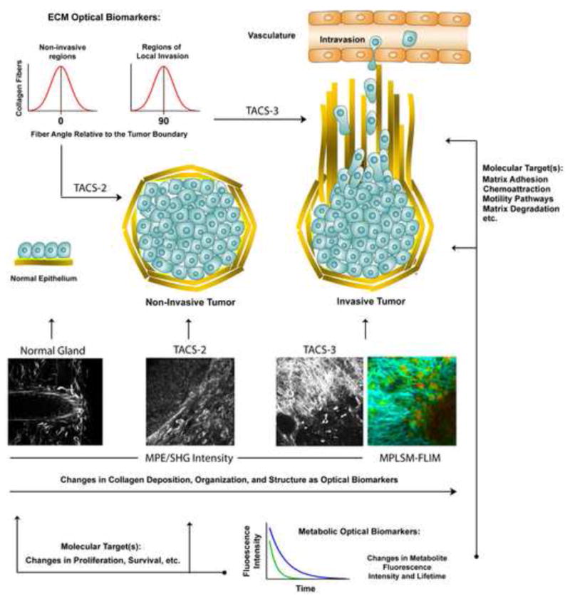
Understanding tissue architecture and physical and chemical reciprocity between cells and their microenvironment provides vital insights into key events in cancer metastasis, such as cell migration through three-dimensional (3D) extracellular matrices. Yet, many mechanistic details associated with metastasis remain elusive due to the difficulty of studying cancer cells in relevant 3D microenvironments. Recently, optical imaging has facilitated the direct observation of single cells as they undertake fundamental steps in the metastatic processes. Optical imaging is also providing novel 'optical biomarkers' with diagnostic potential that are linked to cell-motility pathways associated with metastasis, and these can to help guide new approaches to cancer diagnosis and therapy. Here we present recent advances in one subclass of optical imaging of particular promise for cellular imaging - multiphoton microscopy - that can be used to improve the detection of malignant cells as well as advance our understanding of the cell biology of cancer metastasis.


Conflict of interest statement

Conflict of Interest

The authors declare no conflict of interest.

Figures

Figure 1
Figure 1. Cell phenotypes associated with invasion through the stromal matrix
In vivo MPLSM imaging of tumor cells in mice, as well as MPLSM of tumor cells in relevant 3D matrices, have identified three distinct cell phenotypes: collective invasion, single cell amoeboid migration, and 3D migration of cells with a mesenchymal phenotype. The presence or absence of these phenotypes largely depends on tumor type, the state of the stromal ECM, and the cellular composition of the microenvironment. Collective migration (1), where cells remain physically connected to each other and have a multicellular polarity , can present as sheets of cells with a leading edge, lines of cells or groups of cells, or as a collective group that has detached from the primary tumor. These groups of tumor cells have focalized 3D-matrix adhesions, generate traction force and have an active proteolytic program to modify the ECM. Amoeboid motility (2a) is characterizes by a rounded or ellipsoid morphology, a lack of cell polarity, and non-focalized 3D-matrix adhesions to facilitate high velocity movement through the matrix. Mesenchymal migration (2b) is distinguished by an elongated morphology, focalized 3D-matrix adhesions and an active proteolytic program. Each of these phenotypes are regulated by unique molecular programs. Furthermore, it is clear that invasive cells have multiple compensatory mechanisms to switch between these phenotype in order to efficiently migrate through collagenous matrices. For in depth discussions of these phenotypes the readers in encouraged to examined references ,,.
Figure 2
Figure 2. Optical biomarkers detected with MPLSM
Multiphoton microscopy allows the study of single cell behaviors relevant to metastasis in 3D microenvironments in vitro and in vivo. MPLSM imaging live tumor cells has not only provided new information about the cell biology of metastasis, but has also provided novel optical biomarkers that have diagnostic potential. For instance, changes in the collagen matrix architecture, termed tumor associated collagen signatures (TACS), have been described that help distinguish invasive from non-invasive tumor regions (i.e. TACS-2 vs. TACS-3). Shown are MPLSM micrographs (multiphoton excitation of endogenous fluorescence from FAD with simultaneous SHG of the collagen matrix) of live, intact, mammary gland (i), non-invasive regions of mammary tumor (ii; TACS-2), and invasive regions of mammary tumor (iii; TACS-3; See Box 3). As highlighted in the associated cartoons, the collagen fiber angle distributions associated with these matrix morphologies can be quantified (i.e. fiber angle distribution diagrams shown in the upper left corner), and used to quantitatively distinguish regions of invasion from non-invasive regions of the tumor. Furthermore, changes in cell metabolism that are associated with transformation and tumor progression can be detected in single tumor cells and provide an optical biomarker for disease state (e.g. the MPLSM-FLIM image of TACS-3 showing differences in the fluorescence lifetime of FAD in invading cells versus cells in the primary tumor mass; note the difference in heat map color and intensity in cells in the stroma versus the primary tumor mass). When linked to defined signal transduction pathways, optical biomarkers such as these may help guide therapeutic intervention by indicating whether targeted therapy against a particular pathway is relevant.. Collagen is depicting in yellow and basement membrane in the normal gland is illustrated in light green. MPLSM and MPLSM-FLIM images are adapted and reproduced from Provenzano et al., , with permission.

References

    1. Sahai E. Illuminating the metastatic process. Nat Rev Cancer. 2007;7 (10):737–749. - PubMed
    1. Gupta GP, Massague J. Cancer metastasis: building a framework. Cell. 2006;127 (4):679–695. - PubMed
    1. Massoud TF, Gambhir SS. Molecular imaging in living subjects: seeing fundamental biological processes in a new light. Genes Dev. 2003;17 (5):545–580. - PubMed
    1. Weissleder R. Scaling down imaging: molecular mapping of cancer in mice. Nat Rev Cancer. 2002;2 (1):11–18. - PubMed
    1. Gambhir SS. Molecular imaging of cancer with positron emission tomography. Nat Rev Cancer. 2002;2 (9):683–693. - PubMed

Publication types

Substances