

Fluorescent proteins: a cell biologist's user guide
- PMID: 19819147
- PMCID: PMC2784028
- DOI: 10.1016/j.tcb.2009.08.002
Fluorescent proteins: a cell biologist's user guide
Abstract
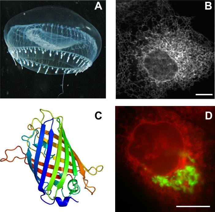
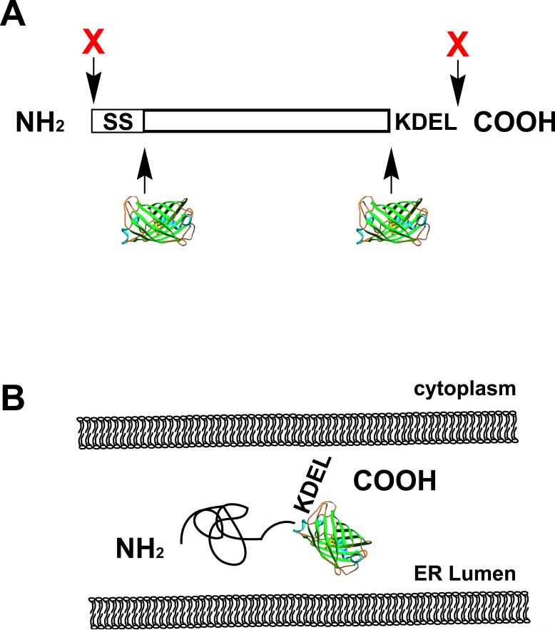
Fluorescent Proteins (FPs) have revolutionized cell biology. The value of labeling and visualizing proteins in living cells is evident from the thousands of publications since the cloning of Green Fluorescent Protein (GFP). Biologists have been flooded with a cornucopia of FPs; however, the FP toolbox has not necessarily been optimized for cell biologists. Common FP plasmids are suboptimal for the construction of proteins fused to FP. More problematic are commercial and investigator-constructed FP-fusion proteins that disrupt important cellular targeting information. Even when cell biologists correctly construct FP-fusion proteins, it is rarely self-evident which FP should be used. Important FP information, such as oligomer formation or photostability, is often obscure or anecdotal. This brief guide is offered to assist the biologist to exploit FPs in the analysis of cellular processes.
Figures
References
-
- Tsien RY. The green fluorescent protein. Annual Review of Biochemistry. 1998;67:509–544. - PubMed
-
- Giepmans BN, et al. The fluorescent toolbox for assessing protein location and function. Science (New York, N.Y. 2006;312:217–224. - PubMed
-
- Shaner NC, et al. Improved monomeric red, orange and yellow fluorescent proteins derived from Discosoma sp. red fluorescent protein. Nature Biotechnology. 2004;22:1567–1572. - PubMed
-
- Lukyanov KA, et al. Innovation: Photoactivatable fluorescent proteins. Nature Reviews. 2005;6:885–891. - PubMed
-
- Lippincott-Schwartz J, Patterson GH. Fluorescent proteins for photoactivation experiments. Methods in Cell Biology. 2008;85:45–61. - PubMed
Publication types
MeSH terms
Substances
Grants and funding
LinkOut - more resources
Full Text Sources
Other Literature Sources
Miscellaneous
