. 2009 Dec 4;284(49):34075-83.
doi: 10.1074/jbc.M109.070235. Epub 2009 Oct 9.

Functional studies of split Arabidopsis Ca2+/H+ exchangers

Affiliations

Functional studies of split Arabidopsis Ca2+/H+ exchangers

Jian Zhao et al. J Biol Chem. .

Abstract

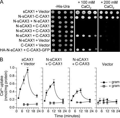
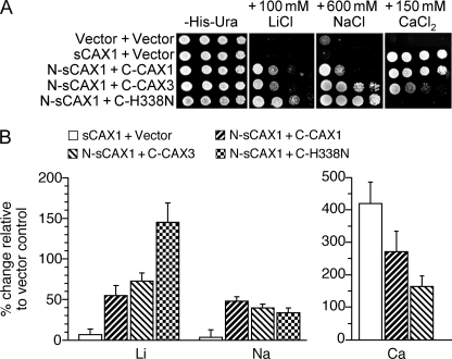
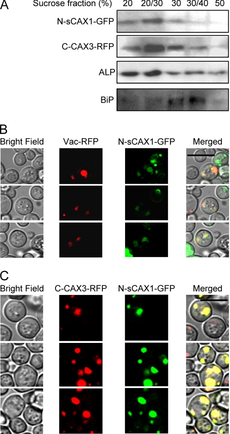
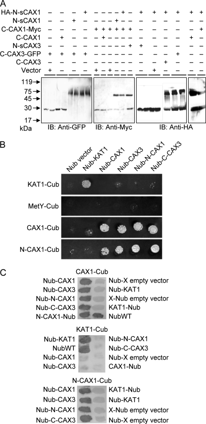
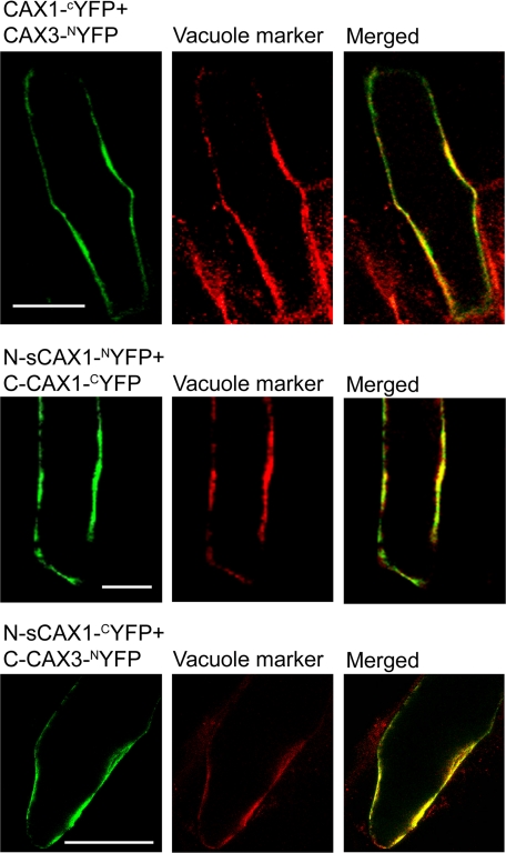
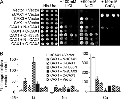
In plants, high capacity tonoplast cation/H(+) antiport is mediated in part by a family of cation exchanger (CAX) transporters. Functional association between CAX1 and CAX3 has previously been shown. In this study we further examine the interactions between CAX protein domains through the use of nonfunctional halves of CAX transporters. We demonstrate that a protein coding for an N-terminal half of an activated variant of CAX1 (sCAX1) can associate with the C-terminal half of either CAX1 or CAX3 to form a functional transporter that may exhibit unique transport properties. Using yeast split ubiquitin, in planta bimolecular fluorescence complementation, and gel shift experiments, we demonstrate a physical interaction among the half proteins. Moreover, the half-proteins both independently localized to the same yeast endomembrane. Co-expressing variants of N- and C-terminal halves of CAX1 and CAX3 in yeast suggested that the N-terminal region mediates Ca(2+) transport, whereas the C-terminal half defines salt tolerance phenotypes. Furthermore, in yeast assays, auto-inhibited CAX1 could be differentially activated by CAX split proteins. The N-terminal half of CAX1 when co-expressed with CAX1 activated Ca(2+) transport, whereas co-expressing C-terminal halves of CAX variants with CAX1 conferred salt tolerance but no apparent Ca(2+) transport. These findings demonstrate plasticity through hetero-CAX complex formation as well as a novel means to engineer CAX transport.


Figures

FIGURE 1.
FIGURE 1.
Functionality of co-expressed CAX half proteins in yeast. A, suppression of Ca2+ sensitivity in K667 yeast through co-expression of various combinations of sCAX1 N-terminal half protein (N-sCAX1), sCAX3 N-terminal half protein (N-sCAX3), CAX1 C-terminal half protein (C-CAX1), and CAX3 C-terminal half protein (C-CAX3), plus epitope-tagged HA-N-sCAX1+C-CAX3-GFP. Saturated liquid cultures of K667 yeast cells were diluted in stepwise 5-fold dilutions and spotted onto selection medium and YPD medium containing either 100 mm or 200 mm CaCl2. Yeast cells expressing sCAX1 and empty vector were used as controls. The plates were incubated and photographed after 2 days at 30 °C. B, time course of 45Ca2+ uptake into endomembrane vesicles prepared from K667 yeast co-expressing sCAX1+empty vector, N-sCAX1+C-CAX1, N-sCAX1+C-CAX3, and empty vector alone. Solid circle, pH-dependent 45Ca2+ uptake; open square, uptake in the presence of the protonophore gramicidin. The Ca2+ ionophore A23187 (5 μm) was added at 12 min. The reduction in Ca2+ uptake following A23187 addition demonstrated that accumulation into the vesicles occurred. The data represent the means ±S.E. of three independent experiments.
FIGURE 2.
FIGURE 2.
Diverse functions of co-expressed CAX half proteins in yeast. A, suppression of Li+, Na+, and Ca2+ sensitivity of K667 yeast cells expressing sCAX1 or co-expressing sCAX1 N-terminal half protein (N-sCAX1) with CAX1 C-terminal half protein (C-CAX1), CAX3 C-terminal half protein (C-CAX3), or CAX1H338N C-terminal half protein (C-H338N). Saturated liquid cultures of K667 yeast were diluted in stepwise 5-fold dilutions and spotted onto selection medium and YPD medium containing 150 mm CaCl2, 100 mm LiCl, or 600 mm NaCl. Yeast cells expressing empty vector were used as a negative control. The plates were incubated and photographed after 2 days at 30 °C. B, Li+, Na+ and Ca2+ ion content analysis in K667 yeast cells co-expressing sCAX1, N-sCAX1+C-CAX1, N-sCAX1+C-CAX3, and N-sCAX1+C-H338N grown to stationary phase in YPD medium supplemented with 500 μm LiCl (for the Li+ and Na+ content measurements) or YPD medium supplemented with 10 mm CaCl2 (for the Ca2+ content measurements). The data from five replicate experiments were calculated with a formula: (Ioncax − Ionvector)/Ionvector × 100, and expressed as the means ± S.E. (n = 5). Ion content varied depending on the growth medium used, but levels of Ca2+ ion content differed among the lines only when the medium was supplemented with CaCl2, whereas Li+ and Na+ content differed among the lines only when the medium was supplemented with LiCl2 and NaCl (data not shown).
FIGURE 3.
FIGURE 3.
Localization of CAX half proteins in yeast cells. Yeast cells expressing GFP-tagged sCAX1 N-terminal half protein (N-sCAX1-GFP) and/or RFP-tagged CAX3 C-terminal half protein (C-CAX3-RFP) were grown overnight in selection medium then diluted with water and observed using sucrose fractionation and confocal microscopy. A, discontinuous sucrose gradient fractionation of microsomes from yeast cells co-expressing N-sCAX1-GFP and C-CAX3-RFP. N-sCAX1-GFP and C-CAX3-RFP were co-fractionated in vacuolar membrane-enriched fractions (20% and 20/30% sucrose interfaces). Alkaline phosphatase (ALP) was used as a vacuolar membrane marker, and BiP was used as an endoplasmic reticulum marker. N-sCAX1-GFP was detected using a GFP antibody and C-CAX3-RFP was detected using an RFP antibody. B and C, co-localization of N-sCAX1-GFP with a prevacuole marker (Vac-RFP) (B) and co-localization of N-sCAX1-GFP and C-CAX3-RFP (C). The panels from left to right are: yeast cells in bright field, RFP image (Vac-RFP or C-CAX3-RFP), GFP image (N-sCAX1-GFP), and merged image. Bar, 10 μm.
FIGURE 4.
FIGURE 4.
Yeast protein interaction assays. A, physical interaction in yeast of N- and C-terminal halves of CAX1 and CAX3 by gel mobility shift assay. The proteins were isolated and solubilized from yeast cells co-expressing HA-tagged sCAX1 N-terminal half (HA-N-sCAX1), sCAX1 C-terminal half (C-CAX1-Myc), or CAX3 C-terminal-GFP fusion (C-CAX3-GFP) with tagged or nontagged sCAX1 N-terminal half (HA-N-sCAX1 or N-sCAX1), CAX1 C-terminal half (C-CAX1-Myc, C-CAX1), CAX3 C-terminal half (C-CAX3 or C-CAX3-GFP), sCAX3 N-terminal half (N-sCAX3), or empty vectors. Following 12% SDS-PAGE and transfer to polyvinylidene difluoride membranes, single tagged split half proteins or protein complexes were detected with immunoblotting (IB) using c-Myc, HA, and GFP antibodies. B, split ubiquitin interaction growth assay. Interaction between CAX1 and CAX3 was determined by growth assay on synthetic minimal medium lacking all six amino acids but supplemented with 150 μm Met for 3 days. The empty Cub and Nub vectors were used as a negative control, and the K+ channel KAT1 was used as a positive control. CAX1-Cub, N-CAX1-Cub, KAT1-Cub, or empty Cub vector (MetY-Cub) were bait for interaction in combination with empty Nub vector, Nub-KAT1, Nub-CAX1, Nub-CAX3, Nub-N-CAX1, and Nub-C-CAX3, respectively. All of the experiments were repeated at least three times with similar results obtained in each replicate. C, X-gal filter assay. Yeast filters set on synthetic minimal medium lacking Ade and His but supplemented with 150 μm Met were grown for 3 days then incubated for 30 min in X-gal solution. All of the experiments were repeated at least three times with similar results obtained in each replicate. The interaction assays are shown with CAX1-Cub (top panel), KAT1-Cub (middle panel), or N-CAX1-Cub (bottom panel) as bait construct and tested in combination with the various Nub-containing prey constructs.
FIGURE 5.
FIGURE 5.
Interaction of CAX half proteins at the tonoplast in plant cells. Onion epidermal cells transiently co-expressing CAX constructs fused to the N-terminal half of YFP (NYFP) and the C-terminal half of YFP (CYFP) (left panels) with a CFP-tagged vacuolar marker (middle panels), and the merged image (right panels). Top row panels, co-expression of CAX1-CYFP and CAX3-NYFP; middle row panels, co-expression of sCAX1 N-terminal half protein (N-sCAX1)-NYFP and CAX1 C-terminal half protein (C-CAX1)-CYFP; bottom row panels, co-expression of N-sCAX1-CYFP and CAX3 C-terminal half protein (C-CAX3)-NYFP. Reconstituted YFP fluorescence signal is shown as false color green, CFP fluorescence signal is shown as false color red, and overlapping YFP/CFP fluorescence signal in the merged image is yellow. Bar, 100 μm.
FIGURE 6.
FIGURE 6.
Functionality of CAX1 and CAX3 when co-expressed with CAX half proteins. A, suppression of Li+, Na+, and Ca2+ sensitivity of K667 yeast cells expressing sCAX1, CAX1, or CAX3, or co-expressing CAX1 with sCAX1 N-terminal half protein (N-sCAX1), CAX1 C-terminal half protein (C-CAX1), sCAX3 N-terminal half protein (N-sCAX3), CAX3 C-terminal half protein (C-CAX3), or CAX1H338N C-terminal half protein (C-H338N). Saturated liquid cultures of K667 yeast were diluted in stepwise 5-fold dilutions and then spotted onto selection medium or YPD medium containing either 150 mm CaCl2, 100 mm LiCl, or 600 mm NaCl. Yeast cells expressing empty vector were used as a negative control. The plates were incubated and photographed after 2 days at 30 °C. B, Li+, Na+, and Ca2+ ion content analysis in K667 yeast cells co-expressing sCAX1 or CAX1 with vector or CAX1 in combination with N-sCAX1, C-CAX1, N-sCAX3, C-CAX3, or C-H338N grown to stationary phase in YPD medium supplemented with 500 μm LiCl (for the Li+ and Na+ content measurements) or 10 mm CaCl2 (for the Ca2+ content measurements). The data from five repeats were calculated with a formula: (Ioncax − Ionvector)/Ionvector × 100, and expressed as the means ± S.E. (n = 5). As discussed previously, the ion content fluctuated depending on the growth medium used.

References

    1. Sanders D., Pelloux J., Brownlee C., Harper J. F. (2002) Plant Cell 14, S401–S417 - PMC - PubMed
    1. Hirschi K. D. (2004) Plant Physiol. 136, 2438–2442 - PMC - PubMed
    1. McAinsh M. R., Pittman J. K. (2009) New Phytol. 181, 275–294 - PubMed
    1. Marty F. (1999) Plant Cell 11, 587–600 - PMC - PubMed
    1. Schumaker K. S., Sze H. (1986) J. Biol. Chem. 261, 2172–2178 - PubMed

Publication types

MeSH terms

LinkOut - more resources