. 2009 Dec 4;284(49):34355-65.
doi: 10.1074/jbc.M109.065730. Epub 2009 Oct 8.

Csm3, Tof1, and Mrc1 form a heterotrimeric mediator complex that associates with DNA replication forks

Affiliations

Csm3, Tof1, and Mrc1 form a heterotrimeric mediator complex that associates with DNA replication forks

Masashige Bando et al. J Biol Chem. .

Abstract

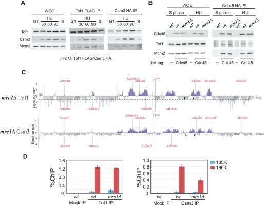
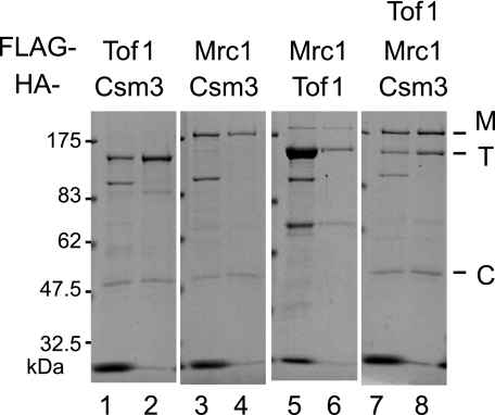
Mrc1 (mediator of replication checkpoint), Tof1 (topoisomerase I interacting factor), and Csm3 (chromosome segregation in meiosis) are checkpoint-mediator proteins that function during DNA replication and activate the effector kinase Rad53. We reported previously that Mrc1 and Tof1 are constituents of the replication machinery and that both proteins are required for the proper arrest and stabilization of replication forks in the presence of hydroxyurea. In our current study, we show that Csm3 is a component of moving replication forks and that both Tof1 and Csm3 are specifically required for the association of Mrc1 with these structures. In contrast, the deletion of mrc1 did not affect the association of Tof1 and Csm3 with the replication fork complex. In agreement with previous observations in yeast cells, the results of a baculovirus coexpression system showed that these three proteins interact directly with each other to form a mediator complex in the absence of replication forks.


Figures

FIGURE 1.
FIGURE 1.
Csm3 is localized at DNA replication forks. A, Tof1 specifically interacts with Mcm2 and Csm3 during S phase. Immunoprecipitations of extracts from wild-type cells that co-expressed Tof1-FLAG3 and Csm3-HA3 were performed using anti-FLAG (FLAG IP) antibodies. Cells were grown exponentially (Asyn.), synchronized in G2 using nocodazole (Noc.), synchronized in G1 using α-factor (G1), and then released from G1 into S phase in the absence (S) or presence (HU) of 200 mm HU for 60 min. Tof1, Csm3, and Mcm2 were then detected by Western blotting analysis of immunoprecipitates (IP) and of a whole cell extract (WCE) control. B, samples were prepared as in A, except in the case of immunoprecipitations using anti-HA antibodies (HA IP). C, distribution of Tof1 and Csm3 on chromosome VI in the wild-type strain in the presence of 200 mm HU. Cells were arrested in G1 by α-factor and then released into S phase at 23 °C in the presence of 200 mm HU for 60 min. Chromatin immunoprecipitation combined with DNA chip hybridization (ChIP-chip) was carried out as described previously (23). Each blue bar represents the binding ratio of Tof1 (upper panel) or Csm3 (lower panel) at each locus and indicates a significant enrichment in some of the IP fractions. The dashed line represents the average signal ratio at the loci that were not enriched (gray bars) in each IP fraction. The scale of the vertical axis is log2. Positions of the centromere (CEN6) and all previously mapped autonomously replicating sequences (ARSs) are shown. The horizontal axis represents the length of chromosome VI in kilobases. Tof1 and Csm3 were tagged with FLAG3 and HA3, respectively. Tof1 and Csm3 were found to localize only in early firing origins of replication (ARS603.5, ARS605, ARS606, and ARS607) under our experimental conditions. D, distribution of Tof1 and Csm3 on chromosome VI in wild-type cells during normal S-phase progression. Cells expressing Tof1-FLAG3 and Csm3-HA3 were arrested in G1 using α-factor and then released into S phase for 40 or 60 min at 16 °C in the presence of Pronase to suppress the rate of DNA synthesis.
FIGURE 2.
FIGURE 2.
Distribution of Cdc45 and rate of BrdUrd incorporation on chromosome VI in wild type (wt) and csm3 deletion mutant yeast in S phase with or without HU. A, distribution of Cdc45 and BrdUrd incorporation on chromosome VI in wild-type and csm3Δ mutant cells in S phase in the presence of HU. Cells expressing a HA-tagged version of Cdc45 were arrested in G1 using α-factor, released into S phase at 23 °C in the presence of 200 mm HU for 60 min, and subjected to ChIP-chip analysis. Blue and red bars represent regions of Cdc45 binding and BrdUrd incorporation, respectively. B, distribution of Cdc45 and BrdUrd incorporation on chromosome VI in wild-type and csm3Δ cells during normal S-phase progression. Cells were arrested in G1 using α-factor, released into S phase for 60 min at 16 °C to slow DNA synthesis, and then processed for ChIP-chip analysis. Blue and red bars represent regions of Cdc45 binding and BrdUrd incorporation, respectively.
FIGURE 3.
FIGURE 3.
Co-dependent association of Tof1 and Csm3 with DNA replication forks. A, interactions among Tof1, Csm3, and Mcm2 were determined by immunoprecipitation using anti-HA antibodies. Extracts of wild-type (wt), tof1Δ, and csm3Δ cells that expressed either Tof1-HA3 or Csm3-HA3 were used in the analysis as indicated. Cells were synchronized in G1 using α-factor and then released into S phase in the absence (S phase) or presence (HU) of 200 mm HU for 60 min. Western blot analyses of Tof1, Csm3, and Mcm2 in immunoprecipitates and in whole cell extract (WCE) controls were performed using anti-HA (for Tof1 and Csm3) and anti-Mcm2 antibodies. B, distribution of Tof1 and Csm3 on chromosome VI in the csm3Δ and tof1Δ mutants under HU-induced replicative stress. Cells were synchronized in G1 using α-factor, released into S phase in the presence of 200 mm HU for 60 min, and processed for ChIP-chip analysis. For a comparison with wild type, see Fig. 1C. C, efficiency of chromatin immunoprecipitation (% ChIP; ratio of input versus IP signal) of Tofl-FLAG3 (left) or Csm3-FLAG3 (right) in wild-type, csm3Δ, and tof1Δ strains was measured by real time PCR analysis using primer pairs specific for amplification of the regions that each begin where indicated by the arrowheads in B; 185K and 196K correspond to the distances in kilobase pairs from the left end of the chromosome. Data represent the average of two independent experiments ± S.D.
FIGURE 4.
FIGURE 4.
Both Tof1 and Csm3 are required for the association of Mrc1 with DNA replication forks. A, interaction between Mrc1 and Mcm2. Extracts from wild-type (wt), tof1Δ, and csm3Δ cells that expressed Mcr1-FLAG3, Tof1-HA3, or Csm3-HA3 were immunoprecipitated using anti-FLAG antibodies. Cells were synchronized in G1 and then released into S phase in the absence (S phase) or presence (HU) of 200 mm HU, followed by Western blotting using anti-FLAG (for Mrc1), anti-HA (for Tof1, Csm3), or anti-Mcm2 antibodies. Whole cell extracts (WCE) were used as a control. B, interactions among Cdc45, Mrc1, and Mcm2. Extracts from wild-type, tof1Δ, csm3Δ, and tof1Δcsm3Δ cells that expressed Cdc45-HA3 and Mcr1-FLAG3 were immunoprecipitated using anti-HA antibodies (HA IP). Cells were synchronized and analyzed as in A. Wild-type cells expressing Mcr1-FLAG3 alone were used as a control (indicated by an asterisk). C, distribution of Mrc1 on chromosome VI in wild-type (wt), tof1Δ, csm3Δ, and tof1Δcsm3Δ cells. Cells were synchronized in G1 using α-factor, released into S phase in the presence of 200 mm HU, and processed for ChIP-chip analysis. Mrc1 distribution was analyzed by ChIP-chip analysis using anti-FLAG antibodies. D, efficiency of chromatin immunoprecipitation (% ChIP; ratio of input versus IP) of Mrc1-FLAG3 in wild-type, tof1Δ, and csm3Δ cells was measured by real time PCR using primer pairs specific for amplification of the regions indicated by the arrowheads in C; 185K and 196K correspond to the distances in kilobase pairs from the left end of the chromosome. Data represent the average of two independent experiments ± S.D.
FIGURE 5.
FIGURE 5.
The association of Tof1 and Csm3 with DNA replication forks is unaffected by Mrc1. A, interaction of Tof1 and Csm3 with Mcm2. Extracts from Δmrc1 cells that co-expressed Tofl-FLAG3 and Csm3-HA3 were immunoprecipitated using anti-FLAG (FLAG IP) or anti-HA (HA IP) antibodies. Cells were arrested in G1 using α-factor (G1), and then released into S phase in the absence (S) or presence (HU) of 200 mm HU for the indicated times (30, 60, or 90 min). Western blotting of immunoprecipitates using anti-FLAG (Tof1), anti-HA (Csm3), and anti-Mcm2 antibodies was then performed. B, interactions among Cdc45, Mcm2, and Tof1 in mrc1Δ and wild-type cells that expressed Cdc45-HA3 and Tofl-FLAG3. Experiments were performed as in A. Wild-type cells that expressed Tofl-FLAG3 alone were used as a control (indicated by an asterisk). C, distribution of Tof1 and Csm3 on chromosome VI in mrc1Δ cells treated with HU. Cell extracts were prepared as described in the legend to Fig. 4B, and ChIP-chip analyses were performed. For a comparison with wild type, see Fig. 1C. D, efficiency of chromatin immunoprecipitation (% ChIP; ratio of input versus IP) of Tofl-FLAG3 and Csm3-FLAG3 in wild-type and mrc1Δ cells was measured by real time PCR using primer pairs specific for amplification of the regions indicated by the arrowheads in C; 185K and 196K correspond to the distances in kilobase pairs from the left end of the chromosome. Data represent the average of two independent experiments ± S.D.
FIGURE 6.
FIGURE 6.
Interactions among Tof1, Csm3, and Mrc1 in SF9 insect cells. Sf9 cells were co-infected with recombinant baculoviruses that expressed untagged, FLAG-tagged or HA-tagged Tof1 (T), HA-tagged Csm3 (C), and FLAG-tagged Mrc1 (M) in the combinations shown. Immunoprecipitation of cell lysates was performed using anti-FLAG M2-agarose beads, and elutions were performed using FLAG peptides (Lanes 1, 3, 5, and 7). The eluate was immunoprecipitated again using anti-HA-agarose beads and eluted with SDS loading buffer without boiling (lanes 2, 4, 6, and 8). Samples were separated by SDS-PAGE and stained with Coomassie Brilliant Blue solution.

References

    1. Branzei D., Foiani M. (2005) Curr. Opin. Cell Biol. 17, 568–575 - PubMed
    1. Lambert S., Carr A. M. (2005) Biochimie 87, 591–602 - PubMed
    1. Labib K., Hodgson B. (2007) EMBO Rep. 8, 346–853 - PMC - PubMed
    1. Tourrière H., Pasero P. (2007) DNA Repair 6, 900–913 - PubMed
    1. Gottifredi V., Prives C. (2005) Semin. Cell Dev. Biol. 16, 355–368 - PubMed

LinkOut - more resources