. 2009 Nov;150(11):5036-45.
doi: 10.1210/en.2009-0771. Epub 2009 Oct 9.

Convergence of 3',5'-cyclic adenosine 5'-monophosphate/protein kinase A and glycogen synthase kinase-3beta/beta-catenin signaling in corpus luteum progesterone synthesis

Affiliations

Convergence of 3',5'-cyclic adenosine 5'-monophosphate/protein kinase A and glycogen synthase kinase-3beta/beta-catenin signaling in corpus luteum progesterone synthesis

Lynn Roy et al. Endocrinology. 2009 Nov.

Abstract

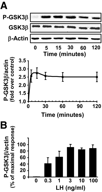
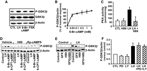
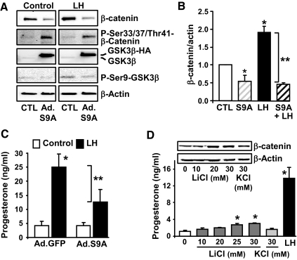
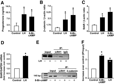
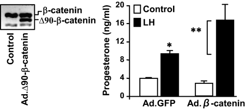
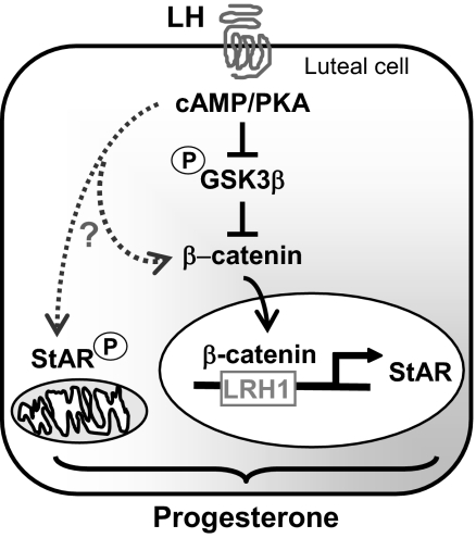
Progesterone secretion by the steroidogenic cells of the corpus luteum (CL) is essential for reproduction. Progesterone synthesis is under the control of LH, but the exact mechanism of this regulation is unknown. It is established that LH stimulates the LH receptor/choriogonadotropin receptor, a G-protein coupled receptor, to increase cAMP and activate cAMP-dependent protein kinase A (PKA). In the present study, we tested the hypothesis that cAMP/PKA-dependent regulation of the Wnt pathway components glycogen synthase kinase (GSK)-3beta and beta-catenin contributes to LH-dependent steroidogenesis in luteal cells. We observed that LH via a cAMP/PKA-dependent mechanism stimulated the phosphorylation of GSK3beta at N-terminal Ser9 causing its inactivation and resulted in the accumulation of beta-catenin. Overexpression of N-terminal truncated beta-catenin (Delta90 beta-catenin), which lacks the phosphorylation sites responsible for its destruction, significantly augmented LH-stimulated progesterone secretion. In contrast, overexpression of a constitutively active mutant of GSK3beta (GSK-S9A) reduced beta-catenin levels and inhibited LH-stimulated steroidogenesis. Chromatin immunoprecipitation assays demonstrated the association of beta-catenin with the proximal promoter of the StAR gene, a gene that expresses the steroidogenic acute regulatory protein, which is a cholesterol transport protein that controls a rate-limiting step in steroidogenesis. Collectively these data suggest that cAMP/PKA regulation of GSK3beta/beta-catenin signaling may contribute to the acute increase in progesterone production in response to LH.


Figures

Figure 1
Figure 1
LH and cAMP stimulate the phosphorylation of GSK3β on ser9 (P-GSK3β) in bovine luteal cells. Primary cultures of steroidogenic luteal cells were treated for various periods of time with LH (0–100 ng/ml). Western blot analysis was performed to determine the levels of GSK3β phosphorylated on ser9 and total GSK3β. β-Actin served as a loading control. A, Time course (0–120 min) of the response to LH (100 ng/ml). A representative Western blot is shown and the graph represents means ± sem from four separate experiments (n = 4). B, Concentration response to LH at 15 min, means ± sem, n = 4.
Figure 2
Figure 2
The stimulatory response of LH on GSK3β phosphorylation (P-GSK3β) is mediated by cAMP/protein kinase (A). Primary cultures of steroidogenic luteal cells were treated as described below. Western blot analysis was performed to determine the levels of total GSK3β and GSK3β phosphorylated on ser9. β-Actin served as a loading control. A, Response to 15 min treatment with control media (CTL), LH (100 ng/ml), 8-Br-cAMP (1 mm), or forskolin (FSK; 10 μm). B, Concentration response to 15 min of treatment with 0.05–2 mm 8-Br-cAMP, means ± sem, n = 4. C, The stimulatory effect of LH on PKA activity is inhibited by the PKA inhibitor H89. Luteal cells were pretreated for 60 min with H89 (20 μm) before treatment for 15 min with LH (100 ng/ml). Protein kinase activity represents means ± sem from three separate experiments. D, PKA inhibitors prevent the stimulatory effects of LH and 8-Br-cAMP on GSK3β phosphorylation. Luteal cells were pretreated for 60 min with the PKA inhibitors H89 (20 μm) or Rp-cAMPS (0.5 μm) and then treated with LH (100 ng/ml), 8-Br-cAMP (1 mm) or 8-(4-chlorophenylthio)-2′-O-methyladenosine-3′, 5′-cAMP (8-CPT-cAMP, 100 μm), a selective activator of EPAC but not PKA. E, Luteal cells were pretreated with the PKC inhibitor GF109203X (GFX; 10 μm), H89, or a combination of inhibitors for 60 min before treatment with LH (100/ml) for 15 min. F, Luteal cells were pretreated for 1 h with the MEK1/2 inhibitor PD98059 (PD; 50 μm) or the phosphatidylinositol 3-kinase inhibitor LY294002 (LY; 20 μm) before treatment with LH for 15 min. Results are means ± sem from three separate experiments.
Figure 3
Figure 3
Modulation of GSK3β activity in luteal cells alters LH-stimulated progesterone secretion. A–C, Luteal cells were infected with an adenovirus-expressing GFP (Ad.GFP) or a hemagglutinin-tagged GSK3β mutant with a ser9 (S9) to alanine mutation (Ad.GSKS9A), which is constitutively active. After 24 h, cells were incubated in the presence or absence of LH (100 ng/ml) for 6 h. A, A representative Western blot panel shows levels of β-catenin, phospho-β-catenin (Ser33/37/Thr41), GSK3β, and phospho (P)-ser9-GSK3β with β-actin as a loading control. B, Levels of total β-catenin protein normalized to actin. *, P < 0.05, control (CTL) vs. Ad.GSKS9A; *, P < 0.05, CTL vs. LH; *, P < 0.05, Ad.GSKS9A vs. LH; **, P < 0.05, LH vs. Ad.GSKS9A+LH. C, Progesterone secretion was measured in aliquots of the media. Results are means ± sem from four separate experiments. **, P < 0.05, Ad.GFP+LH vs. Ad.GSKS9A+LH; *, P < 0.05, Ad.GFP vs. Ad.GFP+LH. D, Effects of LiCl, a GSK3β inhibitor, on progesterone secretion. Luteal cells were treated for 6 h with LiCl (10–30 mm), KCl (30 mm), or LH (100 ng/ml). Shown are representative Western blots of β-catenin and β-actin as a loading control. Progesterone secretion was measured in aliquots of the media. Results are means ± sem from four separate experiments. *, P < 0.05, vs. control.
Figure 4
Figure 4
LH increases β-catenin and StAR in luteal cells. Primary cultures of steroidogenic luteal cells were treated as described below. A–C, Luteal cells were treated with control media or LH (100 ng/ml) or 8 Br-cAMP (1 mm) for 6 h. Results are means ± sem, n = 4 separate experiments. A, Progesterone secretion in response to LH and 8-Br-cAMP. B, β-Catenin protein expressed as a ratio of β-catenin to β-actin. C, StAR levels expressed as a ratio of StAR to β-actin. D, Luteal cells were treated with control media or LH (100 ng/ml) for 2 h. StAR mRNA levels were normalized to glyceraldehyde-3-phosphate dehydrogenase (GAPDH). Results are means ± sem, n = 3. E, Luteal cells were treated with LH (100 ng/ml) or 8-Br-cAMP (1 mm) for 30 min followed by immunoprecipitation (IP) using antibodies to β-catenin or LRH1 or IgG and subsequent ChIP assay showing either LRH1 or β-catenin binding to the proximal StAR promoter. F, Luteal cells were treated with LH (100 ng/ml) or 8-Br-cAMP (1 mm) for 30 min followed by IP of β-catenin and ChIP assay showing β-catenin binding to the proximal StAR promoter. Results are expressed as a ratio of the signal observed in the IP to that of the input. Shown are means ± sem, n = 3 for treatment with 8-Br-cAMP and the averages (bars) and individual values (white circle) from two experiments with LH. *, P < 0.05 vs. control.
Figure 5
Figure 5
β-Catenin enhances LH-stimulated progesterone secretion. Luteal cells were infected the adenoviruses Ad.GFP or Ad.Δ90β-catenin for 24 h before treatment with control media or LH (100 ng/ml) for 6 h. A representative Western blot is shown illustrating overexpression of Ad.Δ90β-catenin in luteal cells. Progesterone secretion was determined from aliquots of conditioned media. Data are means ± sem from three separate experiments. *, P < 0.05, Ad.GFP+LH vs. Ad.GFP+control; **, P < 0.05, Ad.GFP+LH vs. Ad.β-catenin+LH.
Figure 6
Figure 6
Convergence of cAMP/PKA and GSK3/β-catenin signals contributes to progesterone synthesis in luteal cells. Our data provide evidence that LH via a cAMP/PKA mechanism phosphorylates and inhibits GSK3β, leading to the accumulation of β-catenin and the interaction of β-catenin with the StAR promoter, events that play a role in the stimulation of progesterone synthesis. LH, presumably acting via cAMP/PKA, exerts control (?) over other events such as the phosphorylation of StAR, β-catenin, or other cellular mediators to provide optimal stimulation of progesterone synthesis.

References

    1. Davis JS, Rueda BR 2002 The corpus luteum: an ovarian structure with maternal instincts and suicidal tendencies. Front Biosci 7:d1949–d1978 - PubMed
    1. Stouffer RL 2003 Progesterone as a mediator of gonadotrophin action in the corpus luteum: beyond steroidogenesis. Hum Reprod Update 9:99–117 - PubMed
    1. Narko K, Saukkonen K, Ketola I, Bützow R, Heikinheimo M, Ristimäki A 2001 Regulated expression of prostaglandin E(2) receptors EP2 and EP4 in human ovarian granulosa-luteal cells. J Clin Endocrinol Metab 86:1765–1768 - PubMed
    1. Regan JW 2003 EP2 and EP4 prostanoid receptor signaling. Life Sci 74:143–153 - PubMed
    1. Niswender GD 2002 Molecular control of luteal secretion of progesterone. Reproduction 123:333–339 - PubMed

Publication types

MeSH terms