. 2009 Dec;151(4):1918-29.
doi: 10.1104/pp.109.141911. Epub 2009 Oct 9.

The impact of water deficiency on leaf cuticle lipids of Arabidopsis

Affiliations

The impact of water deficiency on leaf cuticle lipids of Arabidopsis

Dylan K Kosma et al. Plant Physiol. 2009 Dec.

Abstract

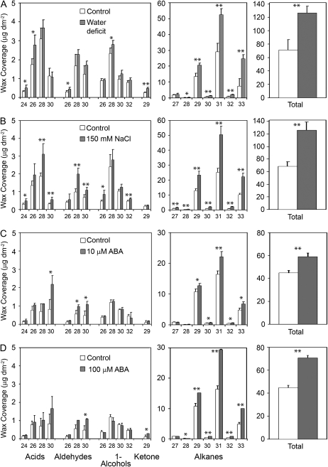
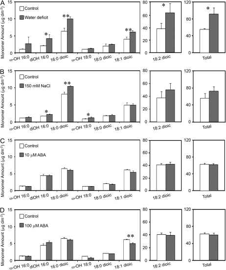
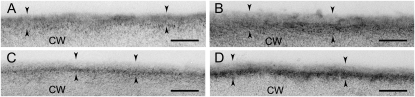
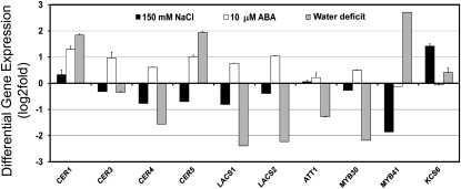
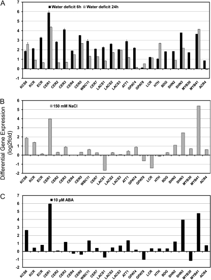
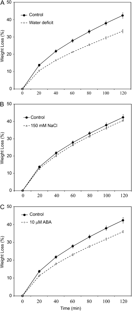
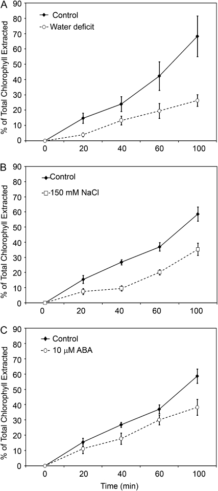
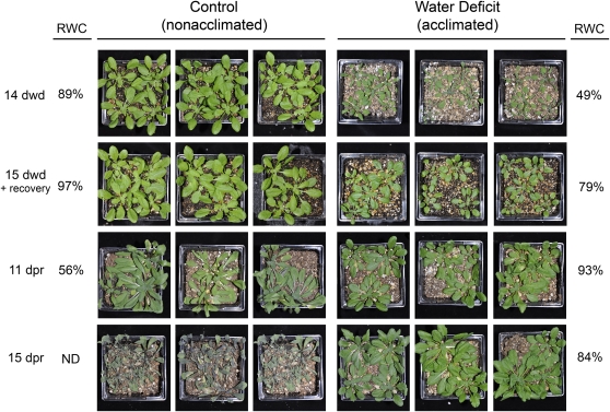
Arabidopsis (Arabidopsis thaliana) plants subjected to water deficit, sodium chloride (NaCl), or abscisic acid treatments were shown to exhibit a significant increase in the amount of leaf cuticular lipids. These stress treatments led to increases in cuticular wax amount per unit area of 32% to 80%, due primarily to 29% to 98% increases in wax alkanes. Of these treatments, only water deficit increased the total cutin monomer amount (by 65%), whereas both water deficit and NaCl altered the proportional amounts of cutin monomers. Abscisic acid had little effect on cutin composition. Water deficit, but not NaCl, increased leaf cuticle thickness (by 49%). Electron micrographs revealed that both water-deprived and NaCl-treated plants had elevated osmium accumulation in their cuticles. The abundance of cuticle-associated gene transcripts in leaves was altered by all treatments, including those performed in both pot-grown and in vitro conditions. Notably, the abundance of the ECERIFERUM1 gene transcript, predicted to function in alkane synthesis, was highly induced by all treatments, results consistent with the elevated alkane amounts observed in all treatments. Further, this induction of cuticle lipids was associated with reduced cuticle permeability and may be important for plant acclimation to subsequent water-limited conditions. Taken together, these results show that Arabidopsis provides an excellent model system to study the role of the cuticle in plant response to drought and related stresses, and its associated genetic and cellular regulation.


Figures

Figure 1.
Figure 1.
Cuticular wax profiles of Arabidopsis rosette leaves from plants subjected to water deficit (A), 150 mm NaCl (B), 10 μm ABA (C), or 100 μm ABA (D). ABA-treated plants and their respective controls were greenhouse grown. Methanol was used to dissolve ABA, which was diluted to treatment concentrations with distilled, deionized water. Control plants for ABA treatments were sprayed with distilled, deionized water containing an equivalent amount of methanol. NaCl-treated, water deficit-treated, and their respective control plants were grown in a growth room. Bars represent mean values ± sd (n = 3–4, the experiments were repeated once, twice in the case of water deficit, with similar results). Asterisks denote significant differences (*, P < 0.05; **, P < 0.01) as determined by Student's t tests or Satterthwaite t tests.
Figure 2.
Figure 2.
Cutin monomer profiles of Arabidopsis rosette leaves from plants subjected to water deficit (A), 150 mm NaCl (B), 10 μm ABA (C), or 100 μm ABA (D). ABA-treated plants and their respective controls were greenhouse grown. Methanol was used to dissolve ABA, which was diluted to treatment concentrations with distilled, deionized water. Control plants for ABA treatments were sprayed with distilled, deionized water containing an equivalent amount of methanol. NaCl-treated, water deficit-treated, and their respective control plants were grown in a growth room. Bars represent mean values ± sd (n = 3–4, the experiments were repeated once with similar results). Asterisks denote significant differences (*, P < 0.05; **, P < 0.01) as determined by Student's t tests or Satterthwaite t tests.
Figure 3.
Figure 3.
Leaf cuticle membrane (between arrowheads) from leaf number four of control plants (A and C), water deficit-treated (B), 150 mm NaCl-treated (D) plants. The cuticle is divided into an outermost, electron-translucent layer, the cuticle proper, and an innermost, electron-dense, darker-staining layer, the cuticular layer, that is most evident in leaves of stressed plants (B and D). CW, Cell wall; scale bars = 100 nm.
Figure 4.
Figure 4.
Stress profiling of cuticle gene transcripts from stress treatments with pot-grown plants. Modulation of cuticle gene expression was determined in rosette leaves of approximately 15-d-old Arabidopsis plants subjected to different stress conditions including water deficit, 150 mm NaCl for 24 h, or 10 μm ABA for 24 h. The gene expression level was determined by quantitative RT-PCR analysis. Results are presented as differential relative transcript abundance. The data represent the means ± sd of three replicates. KCS6 = CER6.
Figure 5.
Figure 5.
Stress profiling of cuticle gene transcripts from in vitro stress treatments. Modulation of cuticle gene expression was determined in 15-d-old Arabidopsis plants subjected to different stress conditions including: water deficit (A), 150 mm NaCl (B), or 10 μm ABA (C). Water deficit treatment consisted of removal of petri dish lids and exposure of plantlets to dry air for 6 or 24 h. All other treatments lasted 24 h. The gene expression level was determined by quantitative RT-PCR analysis. Results are presented as differential relative transcript abundance. The data represent the means ± sd of three replicates. KCS6 = CER6, ECR = CER10.
Figure 6.
Figure 6.
Water-loss rates (expressed as a percentage of initial water-saturated weight) of isolated rosettes from whole plants deprived of water (water deficit; A), subjected to 150 mm NaCl (B), or 10 μm ABA (C). Points represent mean values ± se (n = 5, the experiments were repeated once with similar results).
Figure 7.
Figure 7.
Chlorophyll-extraction rates (expressed as a percentage of total chlorophyll extracted after 24 h) of rosettes from plants deprived of water (water deficit; A), subjected to 150 mm NaCl (B), or 10 μm ABA (C). Points represent mean values ± se (n = 4, the experiments were repeated once with similar results).
Figure 8.
Figure 8.
Water deprivation acclimation experiment. Approximately 15-d-old plants were exposed to 14 d of water deprivation (dwd) until reaching a RWC of 49% alongside well-watered control plants. Both groups of plants were then rewatered and allowed to recover for 20 h. Plants that recovered from water deficit (acclimated) and control plants (nonacclimated) were then subjected to further water deprivation for up to 15 d post recovery (dpr). Water deficit-acclimated plants were resistant to further water deprivation and were able to maintain a higher RWC compared to nonacclimated plants. ND, Not determined.

Similar articles

Cited by

References

    1. Aarts MGM, Keijzer CJ, Stiekema WJ, Pereira A (1995) Molecular characterization of the CER1 gene of Arabidopsis involved in epicuticular wax biosynthesis and pollen fertility. Plant Cell 7 2115–2127 - PMC - PubMed
    1. Barrs HD, Weatherley PE (1962) A re-examination of the relative turgidity technique for estimating water deficits in leaves. Aust J Biol Sci 15 413–428
    1. Bartels D, Sunkar R (2005) Drought and salt tolerance in plants. Crit Rev Plant Sci 24 23–58
    1. Bessire M, Chassot C, Jacquat AC, Humphry M, Borel S, Petétot JC, Métreaux JP, Nawrath C (2007) A permeable cuticle in Arabidopsis leads a strong resistance in Botrytis cinera. EMBO J 26 2158–2168 - PMC - PubMed
    1. Bonaventure G, Beisson F, Ohlrogge J, Pollard M (2004) Analysis of the aliphatic monomer composition of polyesters associated with Arabidopsis epidermis: occurrence of octadeca-cis-6,cis-9-diene-1,18-dioate as the major component. Plant J 40 920–930 - PubMed

Publication types

MeSH terms