

Retinoblastoma protein plays multiple essential roles in the terminal differentiation of Sertoli cells
- PMID: 19819985
- PMCID: PMC2775940
- DOI: 10.1210/me.2009-0184
Retinoblastoma protein plays multiple essential roles in the terminal differentiation of Sertoli cells
Abstract
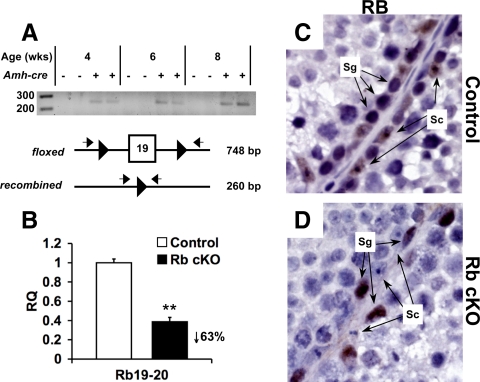
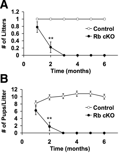
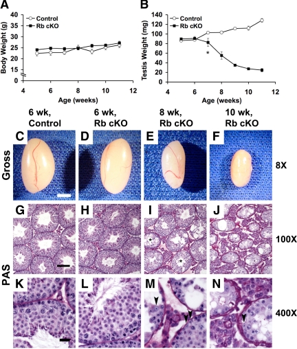
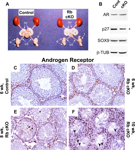
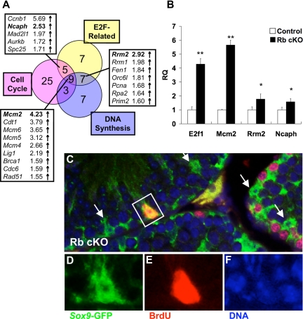
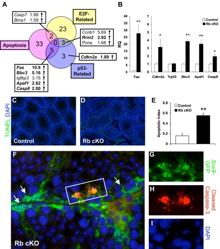
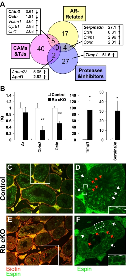
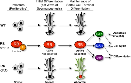
Retinoblastoma protein (RB) plays crucial roles in cell cycle control and cellular differentiation. Specifically, RB impairs the G(1) to S phase transition by acting as a repressor of the E2F family of transcriptional activators while also contributing towards terminal differentiation by modulating the activity of tissue-specific transcription factors. To examine the role of RB in Sertoli cells, the androgen-dependent somatic support cell of the testis, we created a Sertoli cell-specific conditional knockout of Rb. Initially, loss of RB has no gross effect on Sertoli cell function because the mice are fertile with normal testis weights at 6 wk of age. However, by 10-14 wk of age, mutant mice demonstrate severe Sertoli cell dysfunction and infertility. We show that mutant mature Sertoli cells continue cycling with defective regulation of multiple E2F1- and androgen-regulated genes and concurrent activation of apoptotic and p53-regulated genes. The most striking defects in mature Sertoli cell function are increased permeability of the blood-testis barrier, impaired tissue remodeling, and defective germ cell-Sertoli cell interactions. Our results demonstrate that RB is essential for proper terminal differentiation of Sertoli cells.
Figures
Similar articles
-
Absence of inhibin alpha and retinoblastoma protein leads to early sertoli cell dysfunction.PLoS One. 2010 Jul 27;5(7):e11797. doi: 10.1371/journal.pone.0011797. PLoS One. 2010. PMID: 20676395 Free PMC article.
-
Retinoblastoma protein (RB) interacts with E2F3 to control terminal differentiation of Sertoli cells.Cell Death Dis. 2014 Jun 5;5(6):e1274. doi: 10.1038/cddis.2014.232. Cell Death Dis. 2014. PMID: 24901045 Free PMC article.
-
ARID4A and ARID4B regulate male fertility, a functional link to the AR and RB pathways.Proc Natl Acad Sci U S A. 2013 Mar 19;110(12):4616-21. doi: 10.1073/pnas.1218318110. Epub 2013 Mar 4. Proc Natl Acad Sci U S A. 2013. PMID: 23487765 Free PMC article.
-
Retinoblastoma-E2F Transcription Factor Interplay Is Essential for Testicular Development and Male Fertility.Front Endocrinol (Lausanne). 2022 May 19;13:903684. doi: 10.3389/fendo.2022.903684. eCollection 2022. Front Endocrinol (Lausanne). 2022. PMID: 35663332 Free PMC article. Review.
-
Genetic instability as a consequence of inappropriate entry into and progression through S-phase.Cancer Metastasis Rev. 1995 Mar;14(1):59-73. doi: 10.1007/BF00690212. Cancer Metastasis Rev. 1995. PMID: 7606822 Review.
Cited by
-
MicroRNA-449 and microRNA-34b/c function redundantly in murine testes by targeting E2F transcription factor-retinoblastoma protein (E2F-pRb) pathway.J Biol Chem. 2012 Jun 22;287(26):21686-98. doi: 10.1074/jbc.M111.328054. Epub 2012 May 8. J Biol Chem. 2012. PMID: 22570483 Free PMC article.
-
BRG1 Is Dispensable for Sertoli Cell Development and Functions in Mice.Int J Mol Sci. 2020 Jun 19;21(12):4358. doi: 10.3390/ijms21124358. Int J Mol Sci. 2020. PMID: 32575410 Free PMC article.
-
E2F1 controls germ cell apoptosis during the first wave of spermatogenesis.Andrology. 2015 Sep;3(5):1000-14. doi: 10.1111/andr.12090. Andrology. 2015. PMID: 26311345 Free PMC article.
-
UPF2, a nonsense-mediated mRNA decay factor, is required for prepubertal Sertoli cell development and male fertility by ensuring fidelity of the transcriptome.Development. 2015 Jan 15;142(2):352-62. doi: 10.1242/dev.115642. Epub 2014 Dec 11. Development. 2015. PMID: 25503407 Free PMC article.
-
Granulosa cell-expressed BMPR1A and BMPR1B have unique functions in regulating fertility but act redundantly to suppress ovarian tumor development.Mol Endocrinol. 2010 Jun;24(6):1251-66. doi: 10.1210/me.2009-0461. Epub 2010 Apr 2. Mol Endocrinol. 2010. PMID: 20363875 Free PMC article.
References
-
- Skinner MK, Griswold MD 2005 Sertoli cell biology. Boston: Elsevier Academic Press
-
- Vergouwen RP, Jacobs SG, Huiskamp R, Davids JA, de Rooij DG 1991 Proliferative activity of gonocytes, Sertoli cells and interstitial cells during testicular development in mice. J Reprod Fertil 93:233–243 - PubMed
-
- Walker WH 2003 Molecular mechanisms controlling Sertoli cell proliferation and differentiation. Endocrinology 144:3719–3721 - PubMed
-
- Lipinski MM, Jacks T 1999 The retinoblastoma gene family in differentiation and development. Oncogene 18:7873–7882 - PubMed
-
- Nguyen DX, McCance DJ 2005 Role of the retinoblastoma tumor suppressor protein in cellular differentiation. J Cell Biochem 94:870–879 - PubMed
Publication types
MeSH terms
Substances
Grants and funding
LinkOut - more resources
Full Text Sources
Molecular Biology Databases
Research Materials
Miscellaneous
