. 2009 Nov;94(11):4380-90.
doi: 10.1210/jc.2009-0179. Epub 2009 Oct 9.

Impaired fibroblast growth factor receptor 1 signaling as a cause of normosmic idiopathic hypogonadotropic hypogonadism

Affiliations

Impaired fibroblast growth factor receptor 1 signaling as a cause of normosmic idiopathic hypogonadotropic hypogonadism

Taneli Raivio et al. J Clin Endocrinol Metab. 2009 Nov.

Abstract

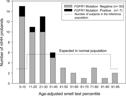
Context: FGFR1 mutations have been identified in about 10% of patients with Kallmann syndrome. Recently cases of idiopathic hypogonadotropic hypogonadism (IHH) with a normal sense of smell (nIHH) have been reported.

Aims: The objective of the study was to define the frequency of FGFR1 mutations in a large cohort of nIHH, delineate the spectrum of reproductive phenotypes, assess functionality of the FGFR1 mutant alleles in vitro, and investigate genotype-phenotype relationships.

Design: FGFR1 sequencing of 134 well-characterized nIHH patients (112 men and 22 women) and 270 healthy controls was performed. The impact of the identified mutations on FGFR1 function was assessed using structural prediction and in vitro studies.

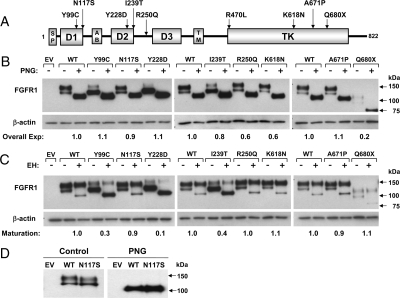
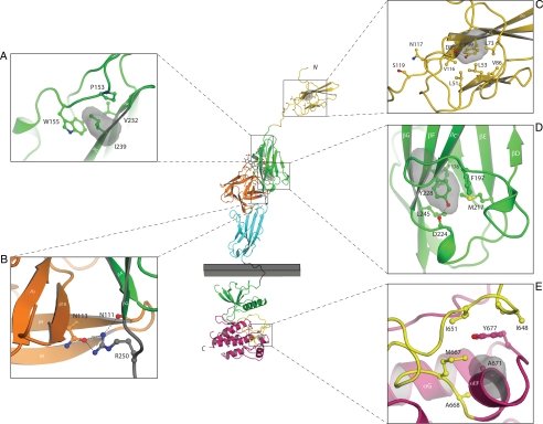
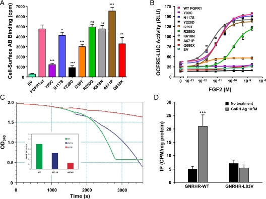
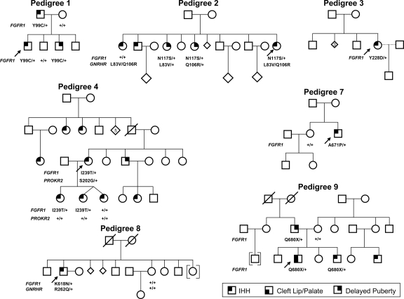
Results: Nine nIHH subjects (five males and four females; 7%) harbor a heterozygous mutation in FGFR1 and exhibit a wide spectrum of pubertal development, ranging from absent puberty to reversal of IHH in both sexes. All mutations impair receptor function. The Y99C, Y228D, and I239T mutants impair the tertiary folding, resulting in incomplete glycosylation and reduced cell surface expression. The R250Q mutant reduces receptor affinity for FGF. The K618N, A671P, and Q680X mutants impair tyrosine kinase activity. However, the degree of functional impairment of the mutant receptors did not always correlate with the reproductive phenotype, and variable expressivity of the disease was noted within family members carrying the same FGFR1 mutation. These discrepancies were partially explained by additional mutations in known IHH loci.

Conclusions: Loss-of-function mutations in FGFR1 underlie 7% of nIHH with different degrees of impairment in vitro. These mutations act in concert with other gene defects in several cases, consistent with oligogenicity.


Figures

Figure 1
Figure 1
A, Schematic of the FGFR1 mutations. The nine FGFR1 mutations span several functional domains of the receptor. AB, Acid box. B–D, Endoglycosidase analysis of FGFR1 mutants. COS-7 cells were transiently transfected with 5 ng of Myc-tagged WT or mutated FGFR1 cDNA. Cell lysates were subjected to PNGase (PNG, B) or EndoH (EH, C) digestion and then processed for FGFR1 immunoblotting using anti-myc antibodies. Overall expression levels were determined by normalizing PNGase-treated bands to their respective β-actin densities (B). Receptor maturation levels were estimated by calculating the fraction of the upper band (mature) out of the total FGFR1 immunoreactivity of EndoH-treated samples (C). In both analyses, the calculated mutant values are expressed as a ratio of WT. Untreated and PNGase-treated WT and N117S mutant are subjected to longer electrophoretic migration (D).
Figure 2
Figure 2
Mapping of the nIHH mutations onto the known FGFR crystal structures suggest that they impair the activity of FGFR1c. The various mutants are mapped onto the ribbon diagram of D1 solution structure (Protein Data Bank entry 2CR3, 2CKN) or FGF2-FGFR1c-heparin complex (Protein Data Bank entry 1FQ9) and FGFR1 kinase domain (Protein Data Bank entry 1FGK) crystal structures. D1 is colored in gold. FGF is colored in orange and the extracellular ligand binding region of FGFR is colored as follows: D2, green; D3, cyan; D2–D3 linker, gray. The coloring of the intracellular tyrosine kinase domain is as follows: the N-terminal lobe of kinase in green; the C-terminal lobe, purple; the activation loop, yellow; and the kinase hinge region, gray. Note that ATP (not shown) binds in the cleft between the N-lobe and C-lobe of the kinase domain. Only relevant β-strands and α-helices of FGFR1c are labeled. A–E, Close-up of the microenvironment of Y99C, N117S, I239, Y228, R250, and A671 subject to mutation in probands with nIHH. In each panel, in addition to the mutated residue, other relevant receptor residues are shown as ball and sticks. A, Side chain of I239 pointing into the hydrophobic core of D2 and interacting with hydrophobic residues P153, W155, and V232, thus contributing to tertiary fold of D2. The surface of I239 is shown as red mesh and P153, W155, and V232 are shown as sticks. B, Network of hydrogen bonds between R250 of FGFR1 and FGF2 is shown. C, Side chain of Y99 pointing into the hydrophobic core of D1 and thus contributing to tertiary fold of D1. The surface of Y99 is shown as red mesh and nearby interacting hydrophobic residues L51, L53, L73, V86, and V116 are shown as sticks. N117 is a potential glycosylation site and is rendered in sticks. D, Side chain of Y228 engages in intramolecular hydrophobic interactions with F176, F197, M217, and L245 in the core of D2. The surface of Y228 is shown as red mesh and F176, F197, M217, and L245 are shown as sticks. E, Surface of A671 is shown in red mesh and the surfaces of hydrophobic residues in the vicinity of A671 are shown in blue mesh. Atom coloring is as follows: nitrogen in blue; oxygen, red; sulfur, yellow. Hydrogen bonds are shown as dashed lines. Letters N and C, N and C termini of FGFR1c, respectively. The membrane bilayer is represented as a gray rectangle.
Figure 3
Figure 3
A, Cell surface expression of FGFR1 mutants. COS-7 cells were transiently transfected with 5 ng of Myc-tagged WT or mutated FGFR1 cDNA. Cell surface receptor levels were assessed using anti-Myc monoclonal antibodies and [125I]rabbit antimouse IgG. Data shown are means ± sem of three experiments, each performed in quadruplicates. Statistical difference between expression of mutants vs. WT receptors were analyzed using repeated-measures ANOVA followed by Dunnett’s multiple comparison test. ns, Not significant. *, P < 0.05; **, P < 0.01; ***, P < 0.001. B, Transcription reporter activity of WT and mutants FGFR1. WT and FGFR1c mutants were transiently transfected into L6 myoblasts with an FGFR1-responsive osteocalcin promoter luciferase construct. FGF2 treatment of WT FGFR1c induced a 5-fold increase in LUC reporter gene expression, whereas Y99C, Y228D, I239T, and Q680X remained silent. The mutant R250Q had a right-shifted curve and did not reach maximum activity of WT. In contrast, N117S, K618N, and A671P activities do not differ from WT. C, Kinase activity of WT and mutants FGFR1. The tyrosine autophosphorylation activity of wild-type (green) and the K621N (blue) and A674P (red) mutant FGFR2 kinases (corresponding to K618N and A671P in FGFR1, respectively) were quantified by using a continuous spectrophotometric assay as described by Barker et al. (43). In this assay, hydrolysis of ATP to ADP is measured as a reduction in NADH absorbance at 340 nm. Relative to wild-type kinase (normalized as 1), the activity of mutant kinases are determined as 0.64 for K621N and 0.25 for A674P consistent with loss of function. D, The L83V hGnRHR mutation blocks GnRH-induced IP accumulation. IP accumulation was measured in COS-7 cells transiently transfected with cDNA encoding wild-type or L83V hGnRHR and stimulated with 10−7 m GnRH agonist. Data points represent the mean ± sem of triplicate samples. The figure is a representative graph from three individual experiments.
Figure 4
Figure 4
Pedigrees of probands with an FGFR1 mutation. Pedigrees 2 and 4 show additional gene defects (GNRHR and PROKR2, respectively). Circles, Females; squares, males; diamonds, additional siblings; arrows, proband; and +, WT for the gene.
Figure 5
Figure 5
Histogram of age-adjusted smell test percentile in nIHH. UPSIT results from seven probands carrying an FGFR1 mutation are contrasted with 50 nIHH probands negative for FGFR1 mutations. The dashed line reflects the number of subjects in the reference population.

Similar articles

Cited by

References

    1. Santoro N, Filicori M, Crowley Jr WF 1986 Hypogonadotropic disorders in men and women: diagnosis and therapy with pulsatile gonadotropin-releasing hormone. Endocr Rev 7:11–23 - PubMed
    1. Seminara SB, Hayes FJ, Crowley Jr WF 1998 Gonadotropin-releasing hormone deficiency in the human (idiopathic hypogonadotropic hypogonadism and Kallmann’s syndrome): pathophysiological and genetic considerations. Endocr Rev 19:521–539 - PubMed
    1. Quinton R, Duke VM, Robertson A, Kirk JM, Matfin G, de Zoysa PA, Azcona C, MacColl GS, Jacobs HS, Conway GS, Besser M, Stanhope RG, Bouloux PM 2001 Idiopathic gonadotrophin deficiency: genetic questions addressed through phenotypic characterization. Clin Endocrinol (Oxf) 55:163–174 - PubMed
    1. Sato N, Katsumata N, Kagami M, Hasegawa T, Hori N, Kawakita S, Minowada S, Shimotsuka A, Shishiba Y, Yokozawa M, Yasuda T, Nagasaki K, Hasegawa D, Hasegawa Y, Tachibana K, Naiki Y, Horikawa R, Tanaka T, Ogata T 2004 Clinical assessment and mutation analysis of Kallmann syndrome 1 (KAL1) and fibroblast growth factor receptor 1 (FGFR1, or KAL2) in five families and 18 sporadic patients. J Clin Endocrinol Metab 89:1079–1088 - PubMed
    1. Kim SH, Hu Y, Cadman S, Bouloux P 2008 Diversity in fibroblast growth factor receptor 1 regulation: learning from the investigation of Kallmann syndrome. J Neuroendocrinol 20:141–163 - PubMed

Publication types

MeSH terms