. 2009 Dec;37(22):7381-93.
doi: 10.1093/nar/gkp833.

DNaseI hypersensitivity at gene-poor, FSH dystrophy-linked 4q35.2

Affiliations

DNaseI hypersensitivity at gene-poor, FSH dystrophy-linked 4q35.2

Xueqing Xu et al. Nucleic Acids Res. 2009 Dec.

Abstract

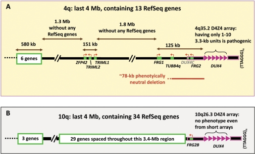
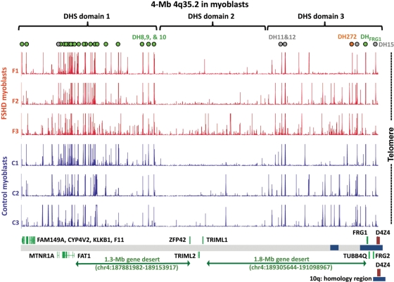
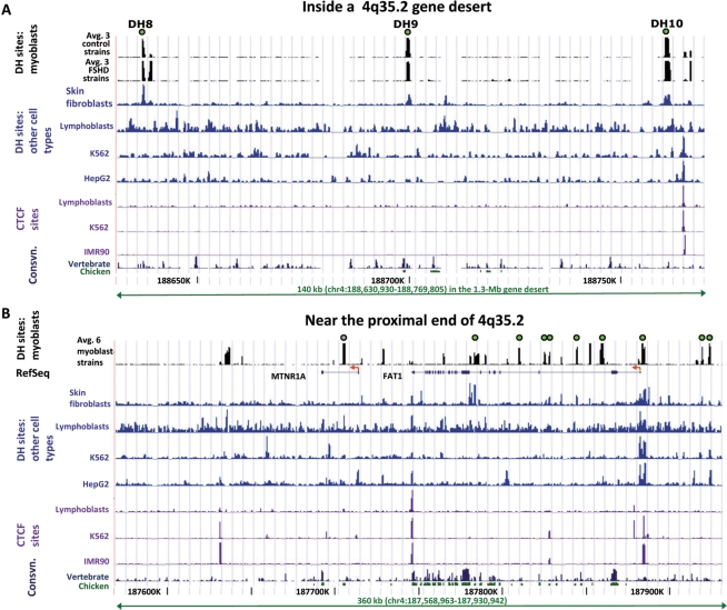
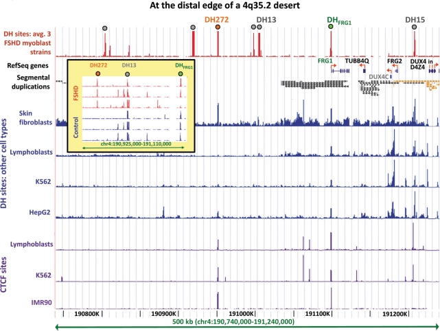
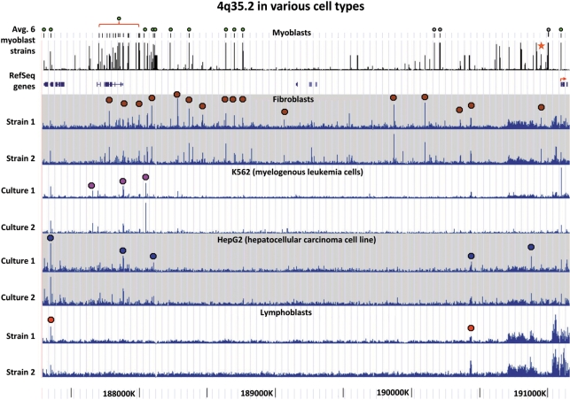
A subtelomeric region, 4q35.2, is implicated in facioscapulohumeral muscular dystrophy (FSHD), a dominant disease thought to involve local pathogenic changes in chromatin. FSHD patients have too few copies of a tandem 3.3-kb repeat (D4Z4) at 4q35.2. No phenotype is associated with having few copies of an almost identical repeat at 10q26.3. Standard expression analyses have not given definitive answers as to the genes involved. To investigate the pathogenic effects of short D4Z4 arrays on gene expression in the very gene-poor 4q35.2 and to find chromatin landmarks there for transcription control, unannotated genes and chromatin structure, we mapped DNaseI-hypersensitive (DH) sites in FSHD and control myoblasts. Using custom tiling arrays (DNase-chip), we found unexpectedly many DH sites in the two large gene deserts in this 4-Mb region. One site was seen preferentially in FSHD myoblasts. Several others were mapped >0.7 Mb from genes known to be active in the muscle lineage and were also observed in cultured fibroblasts, but not in lymphoid, myeloid or hepatic cells. Their selective occurrence in cells derived from mesoderm suggests functionality. Our findings indicate that the gene desert regions of 4q35.2 may have functional significance, possibly also to FSHD, despite their paucity of known genes.


Figures

Figure 1.
Figure 1.
Schematic comparison of the subtelomeric region of 4q containing FSHD-linked D4Z4 arrays (A) and that of 10q, whose D4Z4 arrays are always non-pathogenic (B). Near their distal end, the 4q35.2 and 10q26.3 regions contain ∼98% identical 4q and 10q D4Z4 arrays (8) and other highly homologous sequences outlined in pink. DUX4, within each D4Z4 repeat unit (pink triangle), has no polyA signal except for the most distal copy, which borrows a poly(A) signal from sequences distal to the array (34,35). DUX4 is weakly expressed, has mostly truncated transcripts, is highly polymorphic in number of copies of the repeat (1–100) and shows no more expression from longer than shorter arrays (34,35). Therefore, it was counted as a single gene for the tally of RefSeq genes. DUX4C (grey font) is a predicted gene, not a RefSeq gene.
Figure 2.
Figure 2.
DH sites at 4q35.2 in myoblast cell strains are present in gene deserts as well as in the vicinity of known genes. Green dots, unique DH sites, seen in all six myoblast cultures; orange dot, DH272, seen preferentially in the three FSHD myoblast cell strains and grey dots, DH sites overlapping STRs (70) and seen in all six myoblast cultures. DH site domains are indicated as described in the text. Blue boxes near the bottom of the figure are regions of segmental duplications shared with various chromosomes; the 4q/10q segmental duplication is shown at the bottom of the figure. SLC25A4 (ANT1), which is 5 Mb proximal to D4Z4 and included in the probe set, is not indicated in this figure because it is located on 4q35.1.
Figure 3.
Figure 3.
DH sites in the proximal half of 4q35.2. (A) DH sites 8, 9 and 10 in the 1.3-Mb gene desert of 4q35.2 were seen in myoblasts and fibroblasts but not other cell types. DH sites in the middle of a gene desert are shown for FSHD and control myoblast cell strains and other cell types. For myoblasts, the average data for DH peak height for three FSHD and three control myoblast cultures from DNase-chip are displayed. Individual representative samples from DNase-seq are shown for a skin fibroblast cell strain (GM05879), lymphoblastoid cell line (GM12878), K562 cells (myelogenous leukemia cell line) and HepG2 cells (hepatocellular carcinoma cell line). CTCF binding data for the non-myoblast cells were mapped in IMR90 (lung fibroblasts) by CTCF ChIP following by tiling array analysis (4) and in the other cell types by CTCF ChIP-seq. The single CTCF site in this region is 4 kb distal to DH10. High-resolution mapping of CTCF sites has not yet been done in myoblasts. Consvn, vertebrate (17-way vertebrate alignment) or chicken/human conservation (http://genome.ucsc.edu/). (B) DH sites in the FAT1 and MTNR1A regions are cell type-specific. Tracks and DH sites are labeled as in Panel A.
Figure 4.
Figure 4.
DH272 and DHFRG1 in the terminal 0.4 Mb of 4q of myoblasts overlap DH sites and CTCF sites observed in other cell types. The average data from three FSHD myoblast cell strains are shown; control myoblasts gave the same results except that the DH272 peak was missing or much smaller (inset). Segmental duplications from the UCSC Genome Browser: orange, >99% similarity (mostly to 10q26.3) and grey, 90–98% similarity. Grey dots, DH peaks that overlap STRs (see legend to Figure 2) the only peaks overlapping STRs and seen in all six myoblast cultures in this subregion of 4q35.2 were DH13 and DH15. CTCF data are shown as in Figure 3. DUX4C (grey font) is a predicted gene. Only a few copies of DUX4 within each D4Z4 3.3-kb unit are shown (see Figure 1). The inset displays the mapped DH sites in each of the six studied myoblast cultures in the indicated subregion.
Figure 5.
Figure 5.
Cell type-specific global patterns of DH sites along 4q35.2 even in gene deserts. The average data from three FSHD and three normal control myoblast cultures are shown; green dots, unique-sequence DH sites observed in all six cultures; grey dots, DH sites overlapping STRs and star, DH272. For the other cell types, colored dots indicate the DH peak combination that is most characteristic for a given cell type. The fibroblast tracks are from two skin fibroblast cell strains (GM02185 and GM05879) and the lymphoblast tracks from two lymphoblastoid cell lines (GM19238 and GM19239). The most distal portion of 4q35.2 (∼0.1 Mb in hg18) is not shown because its DH peaks were not reliable, as seen in large variations from sample to sample that we attribute to the many sequences throughout the genome displaying >93% identity to this subregion.
Figure 6.
Figure 6.
Some sequences in 4q35.2 gene deserts near DH sites are transcribed much more than others. (A) Amplicons around DH272 (Supplementary Figure S5) were compared for their average steady-state RNA levels among six myoblast cell strains (pooled data for FSHD and control cell strains, three each) as determined by qRT-PCR. +, distal; −, proximal. P-values (t-test) are given for the comparison of the indicated amplicon to the amplicon 12-kb proximal to DH272. (B) qRT-PCR determinations were done on amplicons located 0.2-kb proximal or distal to the midpoint of the indicated DH site and on the amplicon located 17-kb distal to DH272. P-values are for comparison of the amplicon 17-kb proximal to DH272 to the others.

References

    1. McCann JA, Muro EM, Palmer C, Palidwor G, Porter CJ, Andrade-Navarro MA, Rudnicki MA. ChIP on SNP-chip for genome-wide analysis of human histone H4 hyperacetylation. BMC Genomics. 2007;8:322. - PMC - PubMed
    1. Crawford GE, Holt IE, Whittle J, Webb BD, Tai D, Davis S, Margulies EH, Chen Y, Bernat JA, Ginsburg D, et al. Genome-wide mapping of DNase hypersensitive sites using massively parallel signature sequencing (MPSS) Genome Res. 2006;16:123–131. - PMC - PubMed
    1. Xi H, Shulha HP, Lin JM, Vales TR, Fu Y, Bodine DM, McKay RD, Chenoweth JG, Tesar PJ, Furey TS, et al. Identification and characterization of cell type-specific and ubiquitous chromatin regulatory structures in the human genome. PLoS Genet. 2007;3:e136. - PMC - PubMed
    1. Kim TH, Abdullaev ZK, Smith AD, Ching KA, Loukinov DI, Green RD, Zhang MQ, Lobanenkov VV, Ren B. Analysis of the vertebrate insulator protein CTCF-binding sites in the human genome. Cell. 2007;128:1231–1245. - PMC - PubMed
    1. Barski A, Cuddapah S, Cui K, Roh TY, Schones DE, Wang Z, Wei G, Chepelev I, Zhao K. High-resolution profiling of histone methylations in the human genome. Cell. 2007;129:823–837. - PubMed

Publication types