. 2009 Dec;20(12):2518-24.
doi: 10.1681/ASN.2009030337. Epub 2009 Oct 9.

Th1 and Th17 cells induce proliferative glomerulonephritis

Affiliations

Th1 and Th17 cells induce proliferative glomerulonephritis

Shaun A Summers et al. J Am Soc Nephrol. 2009 Dec.

Abstract

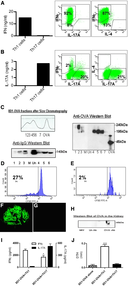
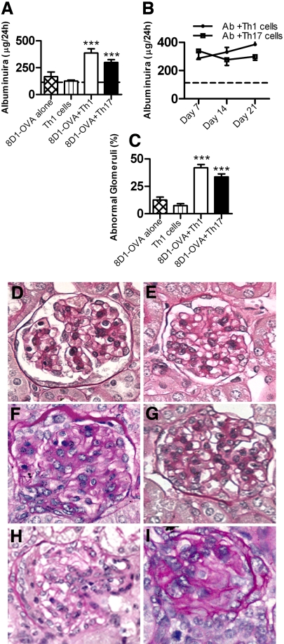
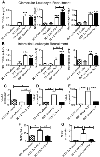
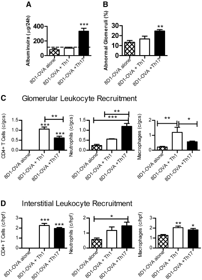
Th1 effector CD4+ cells contribute to the pathogenesis of proliferative and crescentic glomerulonephritis, but whether effector Th17 cells also contribute is unknown. We compared the involvement of Th1 and Th17 cells in a mouse model of antigen-specific glomerulonephritis in which effector CD4+ cells are the only components of adaptive immunity that induce injury. We planted the antigen ovalbumin on the glomerular basement membrane of Rag1(-/-) mice using an ovalbumin-conjugated non-nephritogenic IgG1 monoclonal antibody against alpha3(IV) collagen. Subsequent injection of either Th1- or Th17-polarized ovalbumin-specific CD4+ effector cells induced proliferative glomerulonephritis. Mice injected with Th1 cells developed progressive albuminuria over 21 d, histologic injury including 5.5 +/- 0.9% crescent formation/segmental necrosis, elevated urinary nitrate, and increased renal NOS2, CCL2, and CCL5 mRNA. Mice injected with Th17 cells developed albuminuria by 3 d; compared with Th1-injected mice, their glomeruli contained more neutrophils and greater expression of renal CXCL1 mRNA. In conclusion, Th1 and Th17 effector cells can induce glomerular injury. Understanding how these two subsets mediate proliferative forms of glomerulonephritis may lead to targeted therapies.


Figures

Figure 1.
Figure 1.
Differentiation of OVA-specific OT-II Th1 and Th17 cells, antibody-OVA conjugation, glomerular IgG and intrarenal OVA detection, and recipient immune responses after cell transfer. (A) After stimulating naive OT-II cells with OVA in a Th1 environment, IFNγ was produced and intracellular cytokine staining of CD4+ cells demonstrated strong IFNγ staining with minimal IL-17A or IL-4. (B) Culturing cells in a Th17-stimulating environment led to strong IL-17A production, whereas cells stained positive for IL-17A but not IL-4, and only 2% of cells produced IFNγ. (C) Chromatographic profile of 8D1-OVA conjugation. The numbers 1 to 7 represent fractions collected for analyses by Western blotting, which confirmed that all OVA-conjugated fractions contained OVA and IgG (lanes 1–6), whereas unconjugated fractions (represented as “Un”) contained IgG alone. The lane labeled “M” contained molecular weight markers. (D and E): 8D1-OVA was recognized by OT-II cells because multiple cycles of proliferation of cultured naive OT-II cells (D) were seen with 8D1-OVA conjugate and (E) not seen with unconjugated antibody. Strong linear IgG staining of glomeruli was seen after (F) the administration of 8D1-OVA to Rag1−/− mice, but not after (G) the injection of Th1 cells without antibody. Western blotting of homogenized kidney (H) 24 h after the administration of 8D1-OVA demonstrated OVA in the kidneys (labeled as OVA-Ab); this was not seen after the administration of unconjugated antibody (labeled as Un Ab). (I) Systemic immune responses of recipient Rag1−/− mice at 21 d assessed by splenic cytokine production demonstrated enhanced IFNγ production in mice given 8D1-OVA and Th1 cells, with enhanced IL-17A production by mice receiving 8D1-OVA and Th17 cells. (J) DTH to OVA (at 21 d) was induced only in mice given 8D1-OVA and Th1 cells. *P < 0.05, ***P < 0.001.
Figure 2.
Figure 2.
Renal injury in mice injected with 8D1-OVA conjugate, then either Th1 or Th17 cells. (A) Mice given 8D1-OVA conjugate or Th1 cells alone did not develop albuminuria above values for noninjected Rag1−/− mice (dotted line). At 21 d, albuminuria was increased in mice given 8D1-OVA and Th1 cells or 8D1-OVA and Th17 cells. (B) In mice given 8D1-OVA and Th17 cells, albuminuria had plateaued by day 7 and did not progress. In mice given 8D1-OVA and Th1 cells there was a progressive rise in albuminuria. (C) Histologic injury was significant in mice given 8D1-OVA and either Th1 or Th17 cells. Representative glomeruli from mice given (D) 8D1-OVA alone, (E) Th1 cells alone, (F) 8D1-OVA and Th1 cells, and (G) 8D1-OVA and Th17 cells are shown. (H and I) Crescentic injury and fibrinoid necrosis were only seen in mice given 8D1-OVA and Th1 cells. ***P < 0.001
Figure 3.
Figure 3.
Leukocytes in kidneys of mice with either Th1- or Th17-induced injury 21 d after cell transfer. (A) Glomerular CD4+T cells, neutrophils, and macrophages were increased in mice given 8D1-OVA and Th1/Th17 cells. Neutrophil recruitment was incrementally increased in mice given 8D1-OVA and Th17 cells compared with 8D1-OVA and Th1 cells. (B) A similar pattern of recruitment was seen in the cortical interstitium. Renal chemokine mRNA expression demonstrated (C) enhanced CXCL1 mRNA in mice given 8D1-OVA and Th17 cells, whereas (D) CCL2 and (E) CCL5 were increased in mice given 8D1-OVA and Th1 cells. (F and G) NOS2 and urinary nitrate, markers of macrophage activation, were increased in mice receiving 8D1-OVA and Th1 cells. For mRNA, values for the 8D1-OVA alone group are presented as 1. *P < 0.05, **P < 0.01, ***P < 0.001.
Figure 4.
Figure 4.
Renal disease in mice 3 d after injection with 8D1-OVA and either Th1 or Th17 cells. (A) Pathologic albuminuria (dotted line represents values for noninjected Rag1−/− mice) and (B) increased numbers of abnormal glomeruli were evident in mice that received 8D1-OVA and Th17 cells. (C) Leukocyte recruitment to glomeruli demonstrated CD4+ cells (more in mice receiving Th1 cells), with comparatively more neutrophils in glomeruli of Th17 cell recipients and more macrophages in glomeruli of Th1 cell recipients. (D) Interstitial leukocytes were similar in Th1 and Th17 cell recipients 3 d after cell transfer. *P < 0.05, **P < 0.01, ***P < 0.001.

References

    1. Neale TJ, Tipping PG, Carson SD, Holdsworth SR: Participation of cell-mediated immunity in deposition of fibrin in glomerulonephritis. Lancet 2: 421–424, 1988 - PubMed
    1. Brouwer E, Huitema MG, Mulder AH, Heeringa P, van Goor H, Tervaert JW, Weening JJ, Kallenberg CG: Neutrophil activation in vitro and in vivo in Wegener's granulomatosis. Kidney Int 45: 1120–1131, 1994 - PubMed
    1. Cunningham MA, Huang XR, Dowling JP, Tipping PG, Holdsworth SR: Prominence of cell-mediated immunity effectors in “pauci-immune” glomerulonephritis. J Am Soc Nephrol 10: 499–506, 1999 - PubMed
    1. Huang XR, Tipping PG, Shuo L, Holdsworth SR: Th1 responsiveness to nephritogenic antigens determines susceptibility to crescentic glomerulonephritis in mice. Kidney Int 51: 94–103, 1997 - PubMed
    1. Bolton WK, Chandra M, Tyson TM, Kirkpatrick PR, Sadovnic MJ, Sturgill BC: Transfer of experimental glomerulonephritis in chickens by mononuclear cells. Kidney Int 34: 598–610, 1988 - PubMed

Publication types

MeSH terms