. 2009 Dec;75(23):7343-9.
doi: 10.1128/AEM.01010-09. Epub 2009 Oct 9.

Thermal Stabilization of Erwinia chrysanthemi pectin methylesterase a for application in a sugar beet pulp biorefinery

Affiliations

Thermal Stabilization of Erwinia chrysanthemi pectin methylesterase a for application in a sugar beet pulp biorefinery

Chacko Chakiath et al. Appl Environ Microbiol. 2009 Dec.

Abstract

Directed evolution approaches were used to construct a thermally stabilized variant of Erwinia chrysanthemi pectin methylesterase A. The final evolved enzyme has four amino acid substitutions that together confer a T(m) value that is approximately 11 degrees C greater than that of the wild-type enzyme, while maintaining near-wild-type kinetic properties. The specific activity, with saturating substrate, of the thermally stabilized enzyme is greater than that of the wild-type enzyme when both are operating at their respective optimal temperatures, 60 degrees C and 50 degrees C. The engineered enzyme may be useful for saccharification of biomass, such as sugar beet pulp, with relatively high pectin content. In particular, the engineered enzyme is able to function in biomass up to temperatures of 65 degrees C without significant loss of activity. Specifically, the thermally stabilized enzyme facilitates the saccharification of sugar beet pulp by the commercial pectinase preparation Pectinex Ultra SPL. Added pectin methylesterase increases the initial rate of sugar production by approximately 50%.


Figures

FIG. 1.
FIG. 1.
Summary of the thermally stabilized PME variants after each generation of mutagenesis. The bars indicate the temperature at which 50% of the initial activity was retained after 20 min of heating. The genotypes of the variants are indicated below the bars.
FIG. 2.
FIG. 2.
Results of ruthenium red agar diffusion assay for PME activity. (A) Assay of 3 μl of unheated, induced supernatants from an error-prone PCR library. (B) Assay of 3 μl of induced supernatants heated for 20 min at 53°C. The arrow indicates a candidate mutant at grid position H7; the supernatants at positions C12 and D12 were unheated controls.
FIG. 3.
FIG. 3.
Coupled alcohol oxidase fluorescent assay of induced supernatants from unmutagenized clones and clones from error-prone PCR library. Assay of 15 μl of induced supernatants from unmutagenized, wild-type clones (circles) and mutagenized library clone members (triangles). Each clone's activity was normalized to the mean activity of the distribution of clones for each condition.
FIG. 4.
FIG. 4.
Thermostability of PME variants. Induced supernatants were heated for 20 min at the indicated temperature and were then assayed for initial activity in the AO-coupled fluorescent assay. The initial velocities with the saturating substrate were normalized to the initial velocity of the same unheated, induced supernatant. Closed circles, wild-type; open squares, JL10; ×, JL11; open triangles, JL18; closed diamonds, JL19; open circles, JL25.
FIG. 5.
FIG. 5.
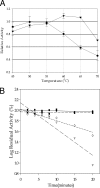
Characterization of the thermal dependence of JL25 PME activity. (A) The relative activity was calculated by subtracting any spontaneous reaction from each enzyme-catalyzed reaction at the corresponding temperature and normalizing each reaction to the activity of the wild-type enzyme at its optimal temperature. Circles, wild type; triangles, JL25. (B) The initial velocities of enzyme heated for the indicated time and at the indicated temperature were determined at 37°C in the coupled AO fluorescent assay, and the activity was normalized to the unheated sample. Circles, JL25 at 62°C; squares, wild type at 50°C; diamonds, JL25 at 65°C; triangles, wild type at 55°C.

References

    1. Abbott, D. W., and A. B. Boraston. 2008. Structural biology of pectin degradation by Enterobacteriaceae. Microbiol. Mol. Biol. Rev. 72:301-316. - PMC - PubMed
    1. Beaulieu, C., M. Boccara, and F. Van Gijsegem. 1993. Pathogenic behavior of pectinase-defective Erwinia chrysanthemi mutants on different plants. Mol. Plant-Microbe Interact. 6:197-202.
    1. Boccara, M., and V. Chatain. 1989. Regulation and role in pathogenicity of Erwinia chrysanthemi 3937 pectin methylesterase. J. Bacteriol. 171:4085-4087. - PMC - PubMed
    1. Bradford, M. M. 1976. A rapid and sensitive method for the quantification of microgram quantities of protein utilizing the principle of protein-dye binding. Anal. Biochem. 72:248-254. - PubMed
    1. Christgau, S., L. V. Kofod, T. Halkier, L. N. Andersen, M. Hockauf, K. Dörreich, H. Dalbøge, and S. Kauppinen. 1996. Pectin methyl esterase from Aspergillus aculeatus: expression cloning in yeast and characterization of the recombinant enzyme. Biochem. J. 319:705-712. - PMC - PubMed

Publication types

MeSH terms

LinkOut - more resources