. 2010 Feb 23;107(8):3299-304.
doi: 10.1073/pnas.0905439106. Epub 2009 Oct 9.

Engineering anatomically shaped human bone grafts

Affiliations

Engineering anatomically shaped human bone grafts

Warren L Grayson et al. Proc Natl Acad Sci U S A. .

Abstract

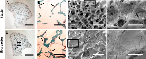
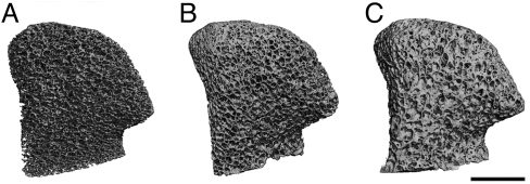
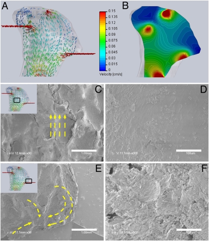
The ability to engineer anatomically correct pieces of viable and functional human bone would have tremendous potential for bone reconstructions after congenital defects, cancer resections, and trauma. We report that clinically sized, anatomically shaped, viable human bone grafts can be engineered by using human mesenchymal stem cells (hMSCs) and a "biomimetic" scaffold-bioreactor system. We selected the temporomandibular joint (TMJ) condylar bone as our tissue model, because of its clinical importance and the challenges associated with its complex shape. Anatomically shaped scaffolds were generated from fully decellularized trabecular bone by using digitized clinical images, seeded with hMSCs, and cultured with interstitial flow of culture medium. A bioreactor with a chamber in the exact shape of a human TMJ was designed for controllable perfusion throughout the engineered construct. By 5 weeks of cultivation, tissue growth was evidenced by the formation of confluent layers of lamellar bone (by scanning electron microscopy), markedly increased volume of mineralized matrix (by quantitative microcomputer tomography), and the formation of osteoids (histologically). Within bone grafts of this size and complexity cells were fully viable at a physiologic density, likely an important factor of graft function. Moreover, the density and architecture of bone matrix correlated with the intensity and pattern of the interstitial flow, as determined in experimental and modeling studies. This approach has potential to overcome a critical hurdle-in vitro cultivation of viable bone grafts of complex geometries-to provide patient-specific bone grafts for craniofacial and orthopedic reconstructions.


Conflict of interest statement

The authors declare no conflict of interest.

Figures

Fig. 1.
Fig. 1.
Tissue engineering of anatomically shaped bone grafts. (A–C) Scaffold preparation. (A and B) Clinical CT images were used to obtain high-resolution digital data for the reconstruction of exact geometry of human TMJ condyles. (C) These data were incorporated into MasterCAM software to machine TMJ-shaped scaffolds from fully decellularized trabecular bone. (D) A photograph illustrating the complex geometry of the final scaffolds that appear markedly different in each projection. (E) The scaffolds were seeded in stirred suspension of hMSCs, to 3 million cells per scaffold (≈1-cm3 volume) and precultured statically for 1 week to allow cell attachment, and then the perfusion was applied for an additional 4 weeks. (F) A photograph of perfusion bioreactor used to cultivate anatomically shaped grafts in vitro. (G–I) Key steps in bioreactor assembly (see Movie S1).
Fig. 2.
Fig. 2.
Cell numbers increased with time of culture and medium perfusion. From day 1 to day 7, the cell numbers increased 7.5-fold, from 3.4 × 10⁶ to 25 × 106 cells per construct. Over the subsequent 4 weeks, cell numbers in static culture increased 4.5-fold to ≈110 × 106 cells per construct, whereas the increase in perfused bioreactor culture was 10-fold to ≈250 × 106 cells per construct (n = 3; *, P < 0.005; **, P < 0.001)
Fig. 3.
Fig. 3.
Bone formation was markedly enhanced by perfusion, in a manner dependent on the fluid flow pattern. (A–D) Constructs cultured under static conditions. (E–H) Constructs cultured with medium perfusion. (A and E) Trichrome staining of entire cross-section of scaffolds showing differences in the new matrix distribution (red) compared with the original scaffold (green) for the static (A) and perfused (E) culture groups. (B and F) Major differences were observed in osteoid formation (arrows) in the central regions of constructs cultured statically (B) and in perfusion (F). (C, D, G, and H) SEM images of the central construct regions. (C and D) Statically cultured constructs exhibit empty pore spaces and loosely packed cells. (G and H) Constructs cultured in perfusion demonstrate formation of dense and confluent lamellae of bone tissue that filled the entire pore spaces. (Scale bars: A and E, 5 mm; B, C, F, and G, 1 mm; D and H, 500 μm.)
Fig. 4.
Fig. 4.
Architecture of the mineralized bone matrix developed with time and in a manner dependent on culture conditions. The reconstructions of 3D μCT images demonstrate the changes in pore structure (relative to the initial state) that were evident at the end of the 5-week cultivation period. Bioreactor constructs exhibit more rapid deposition of new mineral matrix as compared with static constructs (see Table 1). (Scale bar: 5 mm.)
Fig. 5.
Fig. 5.
Bone matrix morphology correlated to the patterns of medium perfusion flow. (A and B) Computational models of medium flow through TMJ constructs during bioreactor cultivation. (A) Color-coded velocity vectors indicate the magnitude and direction of flow through the entire construct based on experimentally measured parameters. (B) Construct is digitally sectioned, and the color-coded contours are used to indicate the magnitude of flow in the inner regions. (C–F) Correlation of the medium flow pattern (by computational modeling) with the structural features of the new bone tissue (by SEM). (C and D) Flat and well-aligned layers of tissue with regular deposition of mineral crystals was observed in the central construct region where flow is unidirectional. (E and F) A swirling flow in a region close to the outlet fluid port resulted in the formation of a swirling tissue structure. (Scale bars: C and E, 1 mm; D and F, 100 μm.)

References

    1. Feinberg SE, Hollister SJ, Halloran JW, Chu TMG, Krebsbach PH. Image-based biomimetic approach to reconstruction of the temporomandibular joint. Cells Tissues Organs. 2001;169:309–321. - PubMed
    1. Grayson W, Chao PHG, Marolt D, Kaplan D, Vunjak-Novakovic G. Engineering custom-designed osteochondral tissue grafts. Trends Biotechnol. 2008;26:181–189. - PMC - PubMed
    1. Alhadlaq A, et al. Adult stem cell driven genesis of human-shaped articular condyle. Ann Biomed Eng. 2004;32:911–923. - PubMed
    1. Alhadlaq A, Mao JJ. Tissue-engineered neogenesis of human-shaped mandibular condyle from rat mesenchymal stem cells. J Dent Res. 2003;82:951–956. - PubMed
    1. Alhadlaq A, Mao JJ. Tissue-engineered osteochondral constructs in the shape of an articular condyle. J Bone Joint Surgery Am. 2005;87:936–944. - PubMed

Publication types

MeSH terms