. 2009 Nov;12(11):1415-23.
doi: 10.1038/nn.2417. Epub 2009 Oct 11.

Presynaptic alpha2delta-3 is required for synaptic morphogenesis independent of its Ca2+-channel functions

Affiliations

Presynaptic alpha2delta-3 is required for synaptic morphogenesis independent of its Ca2+-channel functions

Peri T Kurshan et al. Nat Neurosci. 2009 Nov.

Abstract

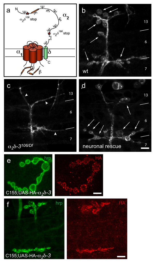
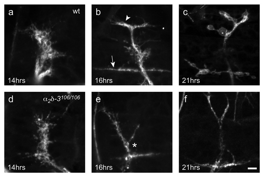
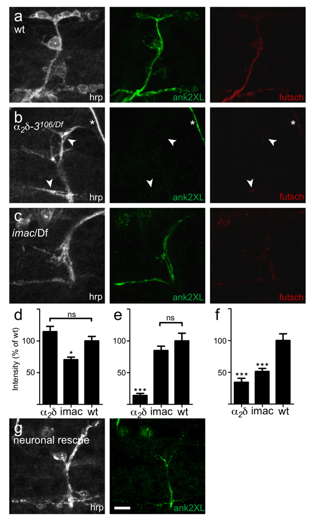
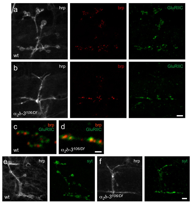
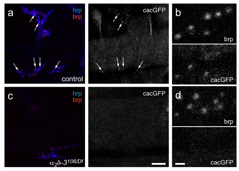
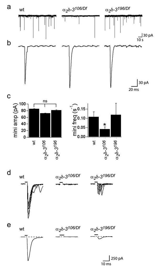
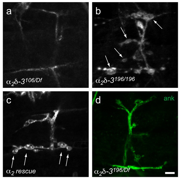
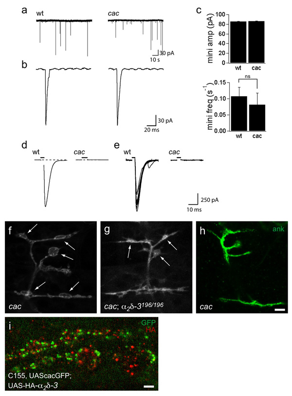
Synaptogenesis involves the transformation of a growth cone into synaptic boutons specialized for transmitter release. In Drosophila embryos lacking the alpha(2)delta-3 subunit of presynaptic, voltage-dependent Ca(2+) channels, we found that motor neuron terminals failed to develop synaptic boutons and cytoskeletal abnormalities arose, including the loss of ankyrin2. Nevertheless, functional presynaptic specializations were present and apposed to clusters of postsynaptic glutamate receptors. The alpha(2)delta-3 protein has been thought to function strictly as an auxiliary subunit of the Ca(2+) channel, but the phenotype of alpha(2)delta-3 (also known as stj) mutations cannot be explained by a channel defect; embryos lacking the pore-forming alpha(1) subunit cacophony formed boutons. The synaptogenic function of alpha(2)delta-3 required only the alpha(2) peptide, whose expression sufficed to rescue bouton formation. Our results indicate that alpha(2)delta proteins have functions that are independent of their roles in the biophysics and localization of Ca(2+) channels and that synaptic architecture depends on these functions.


Figures

Figure 1
Figure 1. α2δ-3 null embryos exhibit a defect in bouton morphogenesis
a. Schematic representation of a voltage-dependent Ca2+ channel, after Arikkath and Campbell. Stars mark the positions of stop codons in the null allele α2δ-3106 and the truncated allele α2δ-3196. The Von Willebrand Factor A (VWFa) homology domain is marked in orange. b–d. Neuronal membranes of embryonic NMJs, 21h AEL, labeled with anti-HRP. b. Wildtype NMJs have rounded boutons (arrows), averaging 17.8 ± 0.6 (SEM, N = 29 NMJs) boutons per A2–A5 hemisegment on embryonic muscles 6, 7, 12 and 13. c. Boutons do not form in α2δ-3106/Df(2R)Ex7128 (arrowheads point to neuronal endings); none were observed in over 100 embryos. d. Neuronal expression of α2δ-3 in the null background rescues bouton formation (arrows; genotype C155elav-gal4/+; α2δ-3106/Df(2R)Ex7128; UAS-HA2δ-3/+). Muscles are indicated by numbers and boundary lines. Scale bar = 5 µm. e, f. HA-tagged α2δ-3 expressed neuronally at a 3rd instar (e) and embryonic (f) NMJs (genotype: C155elav-gal4/+;; UAS-HA2δ-3/+). Scale bars = 5 µm.
Figure 2
Figure 2. α2δ-3 null NMJs exhibit an arrest of synaptic morphogenesis
Developing NMJs of wildtype (a–c) and mutant (d–f) embryos. Neuronal membranes are labeled with anti-HRP. Wildtype motorneuron growth cones reach their target muscles at 13h AEL, – and, as shown at 14h AEL (a), look flat and filamentous. α2δ-3106/106 motorneurons (d) also reach their targets at this time, and appear indistinguishable from wildtype. By 16h AEL, wildtype endings (b) branch in a characteristic pattern, with long neurites extending along the boundary between muscles 6 and 7 (arrow) and a shorter branch forming between muscles 12 and 13 (arrow head). Prevaricosities, still somewhat flat and filamentous, begin to form. At this time, α2δ-3106/106 NMJs are noticeably abnormal, branching in the wrong place (*), lacking neurites along the 12/13 border, and lacking prevaricosities. Just before hatching, wildtype endings (c) have formed rounded boutons but α2δ-3106/106 endings (f) remain as they were at 16h. Scale bar = 5 µm.
Figure 3
Figure 3. Ankyrin2-XL is absent at the α2δ-3 null NMJ
a. A wildtype NMJ immunostained with anti-HRP, anti-ank2-XL and anti-futsch. b. An α2δ-3 null (α2δ-3106/Df(2R)Ex7128) NMJ. Ank2-XL immunostaining was almost completely lacking from these synaptic terminals (arrowheads), and what little did remain was only visible by increasing the gain on the microscope. Loss of ank2-XL staining was specific to synaptic terminals, as wildtype levels of ank2-XL immunoreactivity could be seen in other neuronal compartments, including in the nearby axon of a sensory neuron (asterisk). Futsch staining was also greatly reduced in α2δ-3 mutants. c. imac null mutants, which also lack boutons, nonetheless retained ank-2XL staining. Thus the lack of detectable ank2-XL in α2δ-3 null endings was not simply a result of the morphological abnormality. In comparison, futsch staining was reduced in imac to a similar extent as in α2δ-3. d, e, f. Quantification of staining intensity for hrp (d), ank2-XL (e) and futsch (f). α2δ-3 null mutants exhibited an 86% reduction in ank2-XL immunoreactivity (N = 10 embryos/genotype, p < 0.001, one way ANOVA with Tukey post-hoc test) and a 66% reduction in futsch immunoreactivity (p < 0.001) at the NMJ terminal. imac mutants had no reduction in ank2-XL immunoreactivity, but exhibited a 50% reduction in futsch immunoreactivity (p < 0.001). g. Neuronal expression of α2δ-3 in the mutant background partially restored ank2-XL immunostaining at the endings. Scale bar = 5 µm.
Figure 4
Figure 4. α2δ-3 null embryos retain synaptic components at their terminals
a, b. Wildtype (a) and α2δ-3 null (α2δ-3106/Df(2R)Ex7128) (b) 21h embryos express the active zone protein bruchpilot (anti-brp, red), and glutamate receptors (GluRIIC subunit; green). Neuronal membranes are immunostained with anti-hrp (white). The intensity of individual puncta of BRP did not consistently differ between mutant and wildtype, but α2δ-3106 NMJs had only 45% of the total number of colocalized brp and GluRIIC puncta compared to wildtype (average puncta number ± SEM in wt was 63 ± 8, N = 5 NMJs; in α2δ-3106/Df7128, 29 ± 3, N = 6 NMJs). This reduction parallels the finding that their nerve-muscle contact area was only 39% of that in wildtype NMJs by anti-hrp staining (wt: 160 ± 17 µm2, N = 12 NMJs; α2δ-3106/Df7128: 63 ± 4 µm2, N = 9 NMJs). Moreover, active zones appeared slightly smaller: the total area of colocalized brp and GluRIIC puncta was only 30% of that in wildtype (wt: 14 ± 2 µm2, N = 5 NMJs; α2δ-3106/Df7128: 4.3 ± 0.4 µm2, N = 6 NMJs). c, d. Details of (a) and (b) above; brp and GluRIIC puncta are closely apposed in both wildtype (c) and α2δ-3 (d) embryos. e, f. Synaptotagmin1 (green) is also present in both wildtype (e) and α2δ-3 null (f) embryos. Scale bars = 5 µm (a, b, e, f) and 1 µm (c, d).
Figure 5
Figure 5. Ca2+ channel α1 subunits are not detectable in α2δ-3 null embryonic active zones
a, b. Heterozygous control (Df(2R)Ex7128/+; elavGAL4/UAScacGFP), and c, d. α2δ-3 null (α2δ-3106/Df(2R)Ex7128; elavGAL4;UAScacGFP) NMJs. Anti-HRP staining (blue) marks neuronal membranes and anti-brp (red) labels active zones. Arrows in (a) point to cacGFP fluorescent puncta that colocalize with brp in wildtype. (b) and (d) are enlargements of regions of (a) and (c), respectively. Scale bar = 5 µm in (a, c); 1 µm in (b, d).
Figure 6
Figure 6. α2δ-3 null embryos have spontaneous minis, but no evoked synaptic transmission
Whole-cell patch recordings from embryonic muscles 6 and 7 were performed in voltage-clamp in a modified HL-3 saline containing 1 mM Ca2+ (see Methods). a. Representative traces of spontaneous miniature junctional currents in wildtype, α2δ-3106/106 null and α2δ-3196/196 (“δ-less”) alleles. Average amplitude ± SEM in pA was 85.3 ± 0.6 for wt, 72.0 ± 0.5 for α2δ-3106/106, and 81.5 ± 1.1 for α2δ-3196/196 alleles; N (embryos) = 8 wt, 11 α2δ-3106/106, 4 α2δ-3196/196. Scale bar: 30 pA, 10 s. b. Representative individual minis. Scale bar: 30 pA, 20 ms. c. Mini amplitudes are normal in α2δ-3 mutants, but mini frequency is reduced in α2δ-3106/106 NMJs, from 0.11 ± 0.03 minis/s in wt to 0.04 ± 0.01 minis/s in α2δ-3106/106 nulls. α2δ-3196/196 alleles exhibited normal mini frequency (0.12 ± 0.06 minis/s. p < 0.01, ANOVA with Dunnett posthoc test. d. Overlayed representative evoked responses (10 traces) in 5 mM Ca2+. α2δ-3106/Df(2R)Ex7128 NMJs have no evoked response, while α2δ-3196/Df(2R)Ex7128 NMJs have greatly reduced responses. e. Average evoked responses were obtained from 4 (wildtype), 8 (α2δ-3106/Df(2R)Ex7128), and 4 (α2δ-3196/Df(2R)Ex7128) recordings from different embryos, and 10–40 stimuli per recording. Average amplitude ± SEM in pA was 802 ± 113 for wt, and 76 ± 23 for α2δ-3196/Df(2R)Ex7128; N (embryos) = 4 wt, 4 α2δ-3196/Df(2R)Ex7128. Scale bar = 250 pA, 10 ms in (d, e). Stimulus indicated by black bar above trace.
Figure 7
Figure 7. Bouton formation does not require the δ subunit
Embryonic NMJs on muscles 6, 7, 12, and 13, labeled with anti-HRP (unless otherwise indicated). The α2δ-3 null (α2δ-3106/Df(2R)Ex7128) (a) lacks boutons but they are present (arrows) in the “δ-less” allele (α2δ-3196/196) (b) and when an α2-only construct is expressed in the null mutant background (C155elav-gal4/+; α2δ-3106/Df(2R)Ex7128); UAS-HA-α2/+) (c). d. Ankyrin2-XL is present in the “δ-less” allele. Scale bar = 5 µm.
Figure 8
Figure 8. Bouton formation does not require the cac α1 subunit
a–e. Recordings from wildtype and cac null embryonic NMJs indicate that cac encodes the α1 subunit that mediates evoked transmitter release at embryonic active zones. a. Representative traces depicting spontaneous miniature junctional currents (minis) in wildtype and cacL1320-3 nulls. Scale bar: 30 pA, 10 s. b. Representative individual minis. Scale bar: 30 pA, 20 ms. Mini amplitude and frequency are similar between wildtype and cac nulls. c. Average evoked responses ± SEM in pA was 1051 ± 37 for wildtype in 1 mM Ca2+. No evoked responses are detectable in cac null NMJs. Recordings were obtained from 6 wildtype and 7 cacL13 embryos, with 10–40 stimuli per recording. d. Ten representative evoked responses in 1 mM Ca2+e–g. Synaptic boutons form at NMJs of cac null embryos. NMJs on muscles 6, 7, 12, and 13, labeled with anti-HRP or anti-ank2-XL as indicated. cac nulls (cacL1320–3) (e) develop normal boutons (arrows) and have normal ank2-XL staining (f). cacL1320–3; α2δ-3196/196 double mutants (g) also develop boutons (arrows), although their morphology is slightly abnormal. Scale bar = 5 µm. h. Localization of neuronally expressed cacGFP and HA-tagged α2δ-3. HA-α2δ-3 is not exclusively colocalized at active zones with cacGFP (genotype: C155elav-gal4, UAScacGFP;;UAS-HA-α2δ-3). Scale bar = 2 µm.

References

    1. Lardi-Studler B, Fritschy JM. Matching of pre- and postsynaptic specializations during synaptogenesis. Neuroscientist. 2007;13:115–126. - PubMed
    1. Robitaille R, Adler EM, Charlton MP. Strategic location of calcium channels at transmitter release sites of frog neuromuscular synapses. Neuron. 1990;5:773–779. - PubMed
    1. Wolf M, Eberhart A, Glossmann H, Striessnig J, Grigorieff N. Visualization of the domain structure of an L-type Ca2+ channel using electron cryo-microscopy. J Mol Biol. 2003;332:171–182. - PubMed
    1. Nishimune H, Sanes JR, Carlson SS. A synaptic laminin-calcium channel interaction organizes active zones in motor nerve terminals. Nature. 2004;432:580–587. - PubMed
    1. De Waard M, Gurnett CA, Campbell KP. Structural and functional diversity of voltage-activated calcium channels. Ion Channels. 1996;4:41–87. - PubMed

Publication types

MeSH terms